Class 12 PCM . Chemistry . Chemistry Part II . Biomolecules
Chapter 10: Biomolecules
Ch 10
Chemistry
Class 12 PCM
Charactersits of biomolecules
Biomolecules and Life
- Living systems: They grow, sustain themselves and reproduce through coordinated chemical reactions.
- Biomolecules: Complex molecules such as proteins, nucleic acids and lipids, together with vitamins and mineral salts, support life processes.
Carbohydrates: Definition and Classification
Carbohydrates — Optically active polyhydroxy aldehydes or ketones, or compounds that produce such units on hydrolysis.
Key Features
- Occurrence and examples: Mainly produced by plants; common examples are sucrose, glucose and starch.
- General formula: Many have the form , but this is not a reliable definition: acetic acid fits the formula pattern but is not a carbohydrate, while rhamnose is a carbohydrate but does not fit it.
- Names: Carbohydrates are also called saccharides; sweet carbohydrates are commonly called sugars. Sucrose is cane sugar and lactose is milk sugar.
| Class | Hydrolysis gives | Examples / notes |
|---|---|---|
| Monosaccharides | Cannot be hydrolysed into simpler carbohydrate units | Glucose, fructose, ribose |
| Oligosaccharides | Two to ten monosaccharide units | Includes disaccharides, trisaccharides and tetrasaccharides; sucrose gives glucose and fructose, while maltose gives two glucose units |
| Polysaccharides | A large number of monosaccharide units | Starch, cellulose, glycogen and gums; generally tasteless and called non-sugars |
Reducing and Non-reducing Sugars
- Reducing sugars reduce Fehling’s solution and Tollens’ reagent; all monosaccharides, whether aldoses or ketoses, are reducing sugars.
- Non-reducing sugars do not reduce these reagents; sucrose is an example.
Monosaccharides
| Carbon atoms | General name | Aldehyde group | Ketone group |
|---|---|---|---|
| 3 | Triose | Aldotriose | Ketotriose |
| 4 | Tetrose | Aldotetrose | Ketotetrose |
| 5 | Pentose | Aldopentose | Ketopentose |
| 6 | Hexose | Aldohexose | Ketohexose |
| 7 | Heptose | Aldoheptose | Ketoheptose |
Glucose
Occurrence and Preparation
- Occurrence: Glucose occurs free and combined in nature, especially in sweet fruits, honey and ripe grapes.
- From sucrose: Boiling sucrose with dilute hydrochloric or sulphuric acid in alcoholic solution produces equal amounts of glucose and fructose.
- From starch: Commercial glucose is made by hydrolysing starch with dilute sulphuric acid at under pressure.
Open-chain Structure: Evidence
- Identity: Glucose is an aldohexose, also called dextrose, with molecular formula . It is a monomer of starch and cellulose.
- Straight carbon chain: Prolonged heating with hydroiodic acid forms -hexane, indicating that all six carbon atoms are linked in a straight chain.
- Carbonyl group: Formation of an oxime with hydroxylamine and a cyanohydrin with hydrogen cyanide confirms a carbonyl group.
- Aldehyde group: Bromine water oxidises glucose to gluconic acid, showing that its carbonyl group is aldehydic.
- Five hydroxyl groups: Acetylation produces stable glucose pentaacetate, indicating five hydroxyl groups attached to different carbon atoms.
- Primary alcohol group: Nitric acid oxidises glucose to the dicarboxylic acid saccharic acid; oxidation of gluconic acid also gives saccharic acid, supporting the presence of a primary alcohol group.
The aldehyde end and terminal primary alcohol are shown explicitly.
D/L Configuration and Optical Rotation
How the Configuration Is Assigned
- Reference molecule: D/L configuration in carbohydrates is assigned by comparison with the D- or L-isomer of glyceraldehyde.
- Fischer projection: Write the most oxidised carbon at the top; compare the hydroxyl group on the lowest asymmetric carbon. On the right gives the D configuration; on the left gives the L configuration.
- Glucose: The hydroxyl group on glucose’s lowest asymmetric carbon is on the right, so it is D-glucose.
- Optical rotation is separate: D/L indicates relative configuration, not the direction of rotation. D-glucose is dextrorotatory and is written D-(+)-glucose; D-fructose is laevorotatory.
Do not equate D with (+), or L with (−). D/L describes configuration; (+)/(−) describes measured optical rotation.
Cyclic Structure of Glucose
Why the Open-chain Structure Is Incomplete
- Aldehyde tests: Despite the proposed aldehyde group, glucose does not give Schiff’s test or form a hydrogensulphite addition product with sodium hydrogensulphite.
- Pentaacetate test: Glucose pentaacetate does not react with hydroxylamine, indicating that it has no free aldehyde group.
- Two crystalline forms: Glucose exists as α and β forms, with different melting points and crystallisation conditions; these cannot be explained by the open-chain structure alone.
| Form | Preparation condition | Melting point |
|---|---|---|
| α-glucose | Crystallises from concentrated glucose solution at | |
| β-glucose | Crystallises from hot, saturated aqueous solution at |
Ring Formation and Anomers
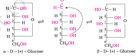
- Hemiacetal formation: The hydroxyl group on C-5 adds to the aldehyde group at C-1, forming a six-membered ring and removing the free aldehyde group.
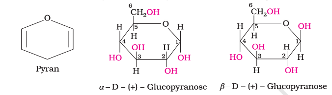
- Anomeric carbon: C-1 becomes the anomeric carbon. The α and β forms differ only in the configuration of the hydroxyl group at this carbon.
- Pyranose: The six-membered glucose ring, containing one oxygen atom and five carbon atoms, is called a pyranose ring.
- Equilibrium: The α and β cyclic forms interconvert through the open-chain form; this interconversion is associated with mutarotation.
Fructose
Structure and Forms
- Identity and occurrence: Fructose is a natural ketohexose with formula . It occurs in fruits, honey and vegetables, and is obtained with glucose by sucrose hydrolysis.
- Open chain: It has a six-carbon straight chain with a ketone group at C-2; it belongs to the D-series but is laevorotatory, so it is D-(−)-fructose.
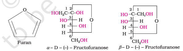
- Cyclic forms: The hydroxyl group at C-5 adds to the C-2 carbonyl group, forming a five-membered ring called a furanose ring.
- Fructose anomers: The α and β forms differ in configuration at the anomeric carbon, which is C-2 in fructose.
Disaccharides
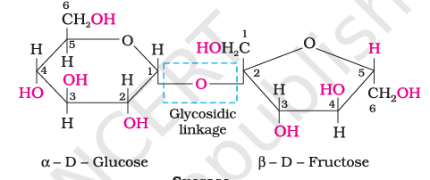
Glycosidic linkage — The oxygen bridge joining two monosaccharide units, formed by loss of a water molecule.
Key Features
- Hydrolysis: Dilute acids or enzymes split a disaccharide into two monosaccharides, which may be identical or different.
- Reducing behaviour: A disaccharide is non-reducing when the reducing groups are involved in the glycosidic bond; a free reducing group makes it reducing.
| Disaccharide | Units and linkage | Reducing property |
|---|---|---|
| Sucrose | Glucose + fructose; hydrolysis gives equimolar D-(+)-glucose and D-(−)-fructose | Non-reducing |
| Maltose | Two α-glucose units; C-1 of one links to C-4 of the other | Reducing: a free aldehyde group can form at C-1 of the second glucose |
| Lactose | β-galactose + β-glucose; C-1 of galactose links to C-4 of glucose | Reducing: glucose can form a free aldehyde group at C-1 |
Invert Sugar
- Rotation change: Sucrose is dextrorotatory, but its hydrolysis mixture is laevorotatory.
- Reason: Fructose’s laevorotation () exceeds glucose’s dextrorotation (), so the mixture rotates light to the left.
- Name: The hydrolysis product is called invert sugar because the sign of rotation changes from positive to negative.
Polysaccharides
Polysaccharides — Polymers containing many monosaccharide units joined by glycosidic linkages; they mainly function in food storage or as structural materials.
| Polysaccharide | Role / occurrence | Structure |
|---|---|---|
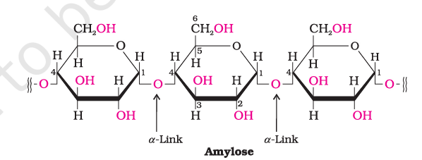
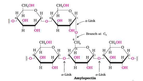
| Starch | Main storage polysaccharide in plants; an important dietary carbohydrate | Polymer of α-glucose, made of amylose and amylopectin |
| Amylose | About 15–20% of starch; water-soluble | Unbranched chain of about 200–1000 α-D-(+)-glucose units; C-1 to C-4 linkages |
| Amylopectin | About 80–85% of starch; water-insoluble | Branched α-glucose polymer; C-1 to C-4 chains and C-1 to C-6 branch points |
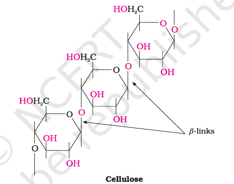
| Cellulose | Plant cell-wall material; the most abundant organic substance in plants | Straight, unbranched chain of β-glucose; C-1 to C-4 linkages |
| Glycogen | Animal carbohydrate store; found in liver, muscles and brain, and also in yeast and fungi | Similar to amylopectin but more highly branched; enzymes break it down to glucose when needed |
Importance of Carbohydrates
- Food and energy: Carbohydrates form a major part of food; honey is a readily available source of energy.
- Storage: Plants store carbohydrates mainly as starch, while animals store them as glycogen.
- Structure and materials: Cellulose forms plant cell walls and is present in wood and cotton; it is used in textiles, paper and related industries.
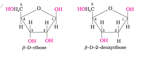
- Nucleic acids: D-ribose and 2-deoxy-D-ribose are aldopentoses found in nucleic acids.
- Biological combinations: Carbohydrates also occur combined with proteins and lipids.
Why are glucose and sucrose soluble in water, while benzene and cyclohexane are not?
What are the products of lactose hydrolysis?
Why does glucose pentaacetate not react with hydroxylamine?
Quick recap
- Carbohydrates are polyhydroxy aldehydes or ketones, or compounds that yield them on hydrolysis.
- Carbohydrates are monosaccharides, oligosaccharides or polysaccharides based on hydrolysis products.
- Glucose is an aldohexose; evidence supports a straight six-carbon chain with an aldehyde, five hydroxyl groups and a primary alcohol group.
- D/L configuration is independent of optical rotation; glucose is D-(+), while fructose is D-(−).
- Glucose forms α- and β-pyranose anomers; fructose forms α- and β-furanose anomers.
- Sucrose is non-reducing; maltose and lactose are reducing disaccharides.
- Starch and glycogen are storage polysaccharides; cellulose is a structural polysaccharide.
Proteins
Proteins and Amino Acids
Protein — A protein is a biomolecule made from one or more chains of α-amino acids linked by peptide bonds.
Importance and sources
- Importance: Proteins are the most abundant biomolecules in living systems and form a basic part of body structure and function.
- Role: They are required for growth and maintenance of the body.
- Sources: Milk, cheese, pulses, peanuts, fish and meat are common protein sources.
Amino Acids
Amino acid — An amino acid contains both an amino group () and a carboxyl group (); proteins yield only α-amino acids on hydrolysis.
Basic structure and names
- General structure: The amino group is attached to the α-carbon, which also bears a side chain : .
- Position-based types: Depending on the amino group's position relative to the carboxyl group, amino acids are called α, β, γ, δ, and so on.
- Side chains: Some amino acids contain additional functional groups in .
- Names and codes: Amino acids have trivial names and are commonly written with three-letter or one-letter codes.
- Name examples: Glycine is named for its sweet taste; tyrosine was first obtained from cheese.
| Amino acid | Side chain | Three-letter | One-letter |
|---|---|---|---|
| Glycine | Gly | G | |
| Alanine | Ala | A | |
| Valine* | Val | V | |
| Leucine* | Leu | L | |
| Isoleucine* | Ile | I | |
| Arginine* | Arg | R | |
| Lysine* | Lys | K | |
| Glutamic acid | Glu | E | |
| Aspartic acid | Asp | D | |
| Glutamine | Gln | Q | |
| Asparagine | Asn | N | |
| Threonine* | Thr | T | |
| Serine | Ser | S | |
| Cysteine | Cys | C | |
| Methionine* | Met | M | |
| Phenylalanine* | Phe | F | |
| Tyrosine | Tyr | Y | |
| Tryptophan* | Indole ring– | Trp | W |
| Histidine* | Imidazole ring– | His | H |
| Proline | Cyclic side chain joins amino N to α-carbon | Pro | P |
Classification and Properties of Amino Acids
| Type | Relative number of groups |
|---|---|
| Neutral | Equal numbers of amino and carboxyl groups |
| Basic | More amino groups than carboxyl groups |
| Acidic | More carboxyl groups than amino groups |
| Point | Essential | Non-essential |
|---|---|---|
| Meaning | Cannot be synthesised by the body; must be obtained from food. | Can be synthesised in the body. |
| Examples from the table | Valine, leucine, isoleucine, arginine, lysine, threonine, methionine, phenylalanine, tryptophan and histidine are marked with *. | The other listed amino acids are not marked with *. |
Physical and ionic properties
- Physical state: Amino acids are generally colourless, crystalline, water-soluble solids with high melting points.
- Reason: They contain both acidic carboxyl and basic amino groups, so they behave like salts rather than simple amines or carboxylic acids.
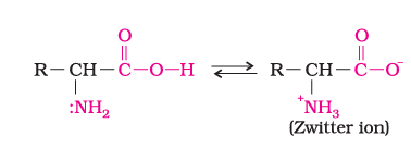
- Zwitterion: In water, the carboxyl group loses a proton and the amino group accepts it, forming a dipolar ion with both charges but no net charge.
- Amphoteric nature: The zwitterionic form can react with both acids and bases.
- Optical activity: Except glycine, naturally occurring α-amino acids are optically active because their α-carbon is asymmetric; they occur as D and L forms.
- L-configuration: Most naturally occurring amino acids are L-amino acids; in the usual representation, the group is on the left.
Structure of Proteins
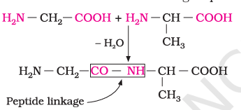
Peptide linkage — A peptide linkage is the amide bond formed between the carboxyl group of one amino acid and the amino group of another.
Peptide formation and chain names
- Condensation: Two amino acids join when one amino group's hydrogen and the other's carboxyl are removed as water.
- Dipeptide: Two amino acids joined by one peptide linkage; glycine plus alanine can form glycylalanine.
- Longer chains: Three, four, five and six residues form tripeptides, tetrapeptides, pentapeptides and hexapeptides, respectively.
- Polypeptide: A chain containing more than ten amino-acid residues.
- Protein: A polypeptide with more than 100 residues and molecular mass above is called a protein; the distinction is not absolute.
- Exception to a simple cutoff: A shorter chain may be called a protein if it has a well-defined protein conformation; insulin has 51 amino acids.
| Type | Chain arrangement and bonds | Solubility | Examples |
|---|---|---|---|
| Fibrous | Polypeptide chains run parallel and are held together by hydrogen and disulphide bonds. | Generally insoluble in water | Keratin (hair, wool, silk); myosin (muscles) |
| Globular | Polypeptide chains coil into a roughly spherical shape. | Usually water-soluble | Insulin; albumins |
Four Levels of Protein Structure
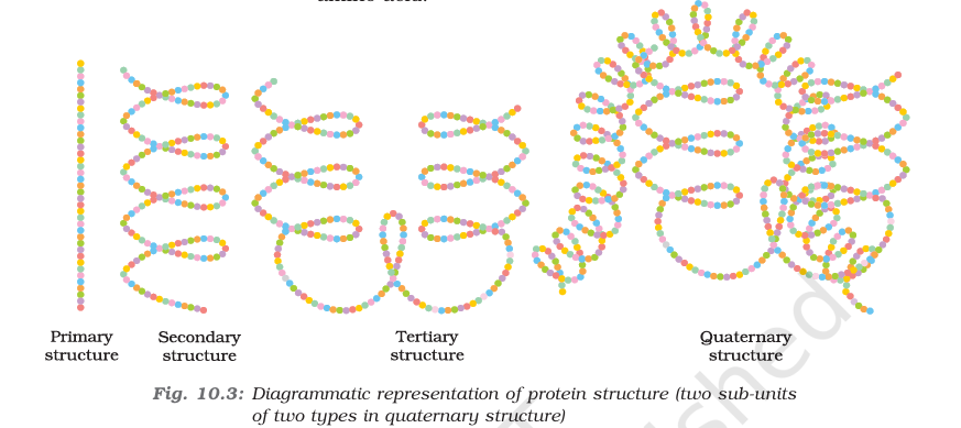
| Level | What it describes | Key feature |
|---|---|---|
| Primary | Specific sequence of amino acids in each polypeptide chain | A change in sequence produces a different protein. |
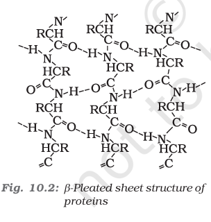
| Secondary | Local shape of a long polypeptide chain | Regular folding caused by hydrogen bonding between peptide-bond and groups. |
| Tertiary | Overall folding of a polypeptide, including further folding of its secondary structure | Produces major fibrous or globular shapes. |
| Quaternary | Spatial arrangement of two or more polypeptide subunits | Present in proteins composed of multiple chains. |
| Point | α-Helix | β-Pleated Sheet |
|---|---|---|
| Arrangement | The chain twists into a right-handed helix. | Stretched chains lie side by side in an extended arrangement. |
| Hydrogen bonding | The group of a residue bonds to the group on an adjacent turn. | Intermolecular hydrogen bonds hold neighbouring chains together. |
| Appearance | Screw-like coil | Pleated, sheet-like folds |
Forces stabilising protein structure
- Stabilising forces: Hydrogen bonds, disulphide linkages, van der Waals forces and electrostatic attractions help stabilise secondary and tertiary structures.
Denaturation of Proteins
Native protein — A native protein has its characteristic three-dimensional structure and biological activity.
Denaturation — Denaturation is the loss of a protein's native shape and biological activity after physical or chemical conditions disrupt its structure.
Denaturation: cause to effect
- Cause: Change in temperature or pH
- Structural effect: Hydrogen bonds are disturbed; globules unfold and helices uncoil.
- Result: Secondary and tertiary structures are destroyed, but the primary structure remains intact.
- Consequence: Biological activity is lost.
Examples
- Boiled egg: Coagulation of egg white is caused by protein denaturation.
- Curdling milk: Lactic acid produced by bacteria causes milk proteins to curdle.
Enzymes and Their Action
Enzyme — An enzyme is a biocatalyst that helps biochemical reactions proceed under the mild conditions found in living organisms.
Enzyme properties
- Composition: Almost all enzymes are globular proteins.
- Specificity: An enzyme acts on a particular substrate and catalyses a particular reaction.
- Naming: Enzymes are generally named after the compound or class of compounds on which they act; maltase hydrolyses maltose to glucose.
- Quantity: Enzymes are needed only in small amounts and, like catalysts, lower activation energy.
| Reaction | Activation energy |
|---|---|
| Acid hydrolysis of sucrose | |
| Hydrolysis by sucrase |
Vitamins
Vitamin — A vitamin is an organic compound needed in small dietary amounts for specific biological functions and normal growth and health.
Key facts
- Need: Vitamin deficiency causes specific diseases; most vitamins cannot be synthesised in the body, although gut bacteria produce some.
- Sources: Plants can synthesise almost all vitamins, and vitamins are generally obtained through the diet.
- Names: Vitamins use letter names such as A, B, C and D; some have subgroups such as , , and .
- Excess: Too much vitamin intake can also be harmful; vitamin pills should not be taken without medical advice.
- Term origin: “Vitamine” was coined from “vital” and “amine”; the final “e” was dropped when most were found not to contain amino groups.
| Group | Vitamins | Solubility and storage |
|---|---|---|
| Fat-soluble | A, D, E, K | Soluble in fats and oils but not water; stored in liver and adipose tissue. |
| Water-soluble | B group and C | Readily excreted in urine and generally not stored, so regular dietary supply is needed; vitamin is an exception. |
| Vitamin | Sources | Deficiency effect |
|---|---|---|
| A | Fish liver oil, carrots, butter and milk | Xerophthalmia (hardening of the cornea); night blindness |
| (thiamine) | Yeast, milk, green vegetables and cereals | Beri beri; loss of appetite and retarded growth |
| (riboflavin) | Milk, egg white, liver and kidney | Cheilosis (fissures at mouth corners and lips), digestive disorders, burning skin sensation |
| (pyridoxine) | Yeast, milk, egg yolk, cereals and grams | Convulsions |
| Meat, fish, egg and curd | Pernicious anaemia (red blood cells deficient in haemoglobin) | |
| C (ascorbic acid) | Citrus fruits, amla and green leafy vegetables | Scurvy (bleeding gums) |
| D | Sunlight exposure, fish and egg yolk | Rickets in children; osteomalacia (soft bones and joint pain) in adults |
| E | Vegetable oils, including wheat germ and sunflower oils | Increased fragility of red blood cells and muscular weakness |
| K | Green leafy vegetables | Increased blood-clotting time |
Why do amino acids generally have higher melting points and greater water solubility than the corresponding halo acids?
What structural changes occur during protein denaturation?
How does an enzyme affect the activation energy of a reaction?
Quick recap
- Proteins: Polymers of α-amino acids joined by peptide linkages; they support body structure, function, growth and maintenance.
- Amino acids: Classified by relative amino/carboxyl group numbers and by whether the body can synthesise them; in water they form zwitterions.
- Protein structures: Primary is sequence, secondary is local folding, tertiary is overall chain folding and quaternary is subunit arrangement.
- Denaturation: Physical or chemical changes disrupt secondary and tertiary structure, not the primary sequence, and remove biological activity.
- Enzymes: Mostly globular protein biocatalysts; they are specific and lower activation energy.
- Vitamins: Small dietary requirements; fat-soluble vitamins are A, D, E, K, while B-group and C are water-soluble.
Nuclear acid
Nucleic Acids: Components and Structure
Nucleic acid — A nucleic acid is a long-chain polymer of nucleotides, involved in transmitting hereditary information.
Main Components
- Chromosomes: Located in the cell nucleus, they contain proteins and nucleic acids and are responsible for heredity.
- Types: The two main nucleic acids are deoxyribonucleic acid (DNA) and ribonucleic acid (RNA).
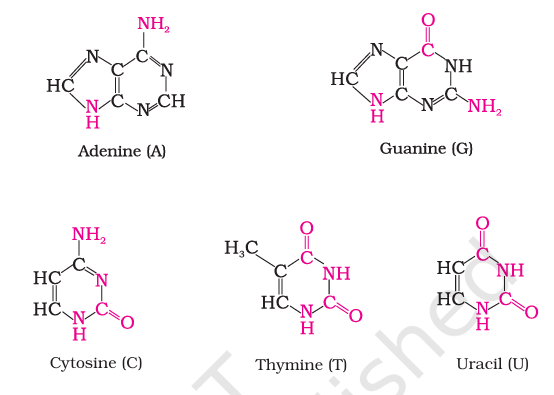
- Complete hydrolysis: DNA or RNA yields a pentose sugar, phosphoric acid and nitrogen-containing bases.
| Feature | DNA | RNA |
|---|---|---|
| Sugar | -D-2-deoxyribose | -D-ribose |
| Bases | Adenine, guanine, cytosine and thymine | Adenine, guanine, cytosine and uracil |
| Usual structure | Double-stranded | Single-stranded |
Nucleosides, Nucleotides and Nucleic-Acid Chains
| Unit | Components |
|---|---|
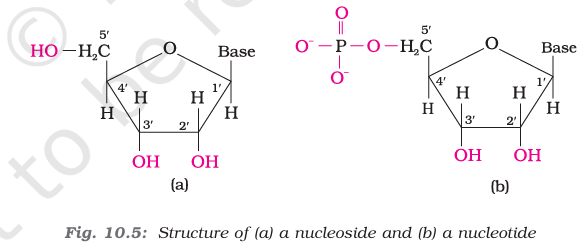
| Nucleoside | Nitrogenous base attached to a pentose sugar |
| Nucleotide | Nucleoside with phosphoric acid attached at the sugar’s position |
- Nucleoside bond: The base attaches to the sugar’s position.
- Sugar numbering: Sugar carbons are labelled , , , and so on, distinguishing them from atoms in the base.
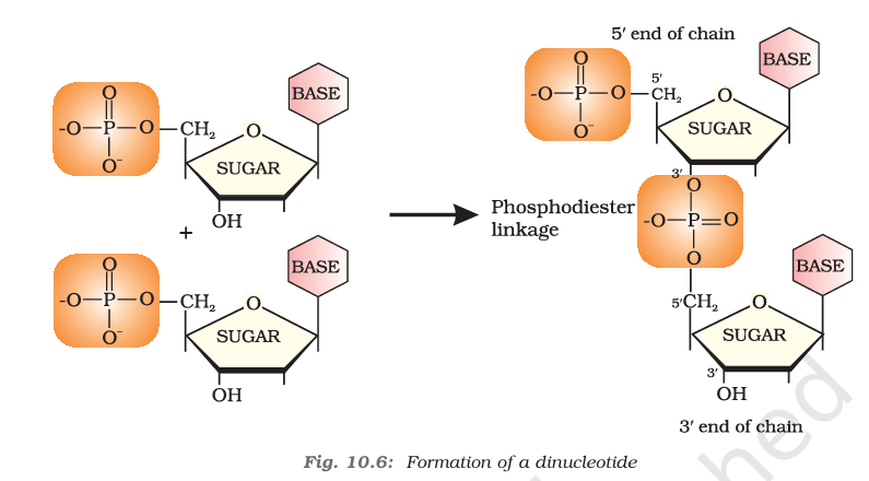
- Phosphodiester linkage: Nucleotides join between the and carbon positions of the pentose sugars.
- Backbone: Repeating sugar–phosphate units form the chain, with bases attached to the sugars.

Primary and Secondary Structure
Primary structure — The primary structure of a nucleic acid is the sequence of nucleotides in its chain.
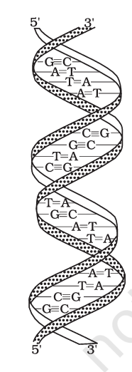
- DNA secondary structure: Watson and Crick described DNA as two chains wound around each other in a double helix.
- Complementary strands: Hydrogen bonds between specific base pairs hold the two strands together.
- Base pairing: Adenine pairs with thymine (A–T); cytosine pairs with guanine (C–G).
- RNA: Hydrolysed RNA bases do not show fixed quantity relationships, consistent with RNA being single-stranded rather than having paired complementary strands.
DNA Fingerprinting
DNA fingerprinting — DNA fingerprinting uses an individual's distinctive sequence of DNA bases for identification.
- Uniqueness: DNA base sequences are individual-specific and the DNA pattern is the same in every cell of that individual.
- Stability: Unlike fingertip patterns that may be altered by surgery, DNA patterns cannot be altered by any known treatment.
- Uses: Criminal identification in forensic laboratories; paternity testing; identifying accident victims by comparison with parents’ or children’s DNA; and identifying racial groups to study biological evolution.
Biological Functions of Nucleic Acids
- DNA and heredity: DNA is the chemical basis of heredity and a reserve of genetic information; it helps maintain species identity over generations.
- Replication: DNA can duplicate during cell division, and identical DNA strands are transferred to daughter cells.
- Protein synthesis: DNA carries the message for making a particular protein; various RNA molecules carry out protein synthesis in the cell.
- RNA types: Messenger RNA (mRNA), ribosomal RNA (rRNA) and transfer RNA (tRNA) are involved in protein synthesis.
Hormones
Hormone — A hormone is an intercellular messenger produced by an endocrine gland and carried in the bloodstream to its site of action.
| Class | Examples |
|---|---|
| Steroids | Estrogens and androgens |
| Polypeptides | Insulin and endorphins |
| Amino-acid derivatives | Epinephrine and norepinephrine |
Hormone Functions
- Regulation: Hormones help maintain the balance of biological activities in the body.
- Blood glucose: Insulin is released when blood glucose rises rapidly and helps keep it within a narrow range; glucagon tends to increase blood glucose.
- External stimuli: Epinephrine and norepinephrine mediate responses to external stimuli.
- Growth and development: Growth hormones and sex hormones have roles in growth and development.
Thyroxine and Thyroid Disorders
Thyroxine — Thyroxine is an iodinated derivative of the amino acid tyrosine, produced by the thyroid gland.
| Condition | Effect |
|---|---|
| Low thyroxine | Hypothyroidism, characterised by lethargy and obesity |
| High thyroxine | Hyperthyroidism |
| Low dietary iodine | May cause hypothyroidism and enlargement of the thyroid gland |
- Iodised salt: Adding sodium iodide to commercial table salt helps control iodine-deficiency disorders.
Adrenal and Gonadal Steroid Hormones
- Sources: Steroid hormones are produced by the adrenal cortex and gonads (testes in males; ovaries in females).
- Glucocorticoids: Control carbohydrate metabolism, modulate inflammatory reactions and help the body respond to stress.
- Mineralocorticoids: Control the excretion of water and salt by the kidneys.
- Addison’s disease: Adrenal-cortex dysfunction may cause hypoglycemia, weakness and increased susceptibility to stress; untreated disease can be fatal. Treatment involves glucocorticoids and mineralocorticoids.
| Hormone | Main role |
|---|---|
| Testosterone | Major male sex hormone; promotes secondary male characteristics such as deep voice, facial hair and general physical constitution |
| Estradiol | Main female sex hormone; promotes secondary female characteristics and participates in menstrual-cycle control |
| Progesterone | Prepares the uterus for implantation of a fertilised egg |
How do a nucleoside and a nucleotide differ?
What products form when a DNA nucleotide containing thymine is completely hydrolysed?
What does the lack of fixed base-quantity relationships in hydrolysed RNA suggest?
Quick recap
- Nucleic acids are nucleotide polymers; complete hydrolysis yields pentose sugar, phosphoric acid and nitrogenous bases.
- DNA has 2-deoxyribose and thymine; RNA has ribose and uracil. Both contain adenine, guanine and cytosine.
- Nucleosides contain a base and sugar; nucleotides also contain phosphate, and polymer chains use phosphodiester linkages.
- DNA is a double helix with complementary A–T and C–G pairs; RNA is generally single-stranded.
- DNA stores and copies hereditary information; DNA fingerprinting is used for identification and paternity testing.
- Hormones are blood-transported messengers; insulin and glucagon regulate blood glucose, while thyroid, adrenal and gonadal hormones have distinct roles.