NEET UG . NEET Zoology . Zoology . Excretory Products and Their Elimination
Chapter 4: Excretory Products and their Elimination
Ch 4
NEET Zoology
NEET UG
Urine Formation
Excretion — Removal of metabolic wastes from the body.
Excretory wastes
- Wastes removed: Animals eliminate ammonia, urea, uric acid, carbon dioxide, water and ions such as , , , phosphate and sulphate.
- Nitrogenous wastes: The main forms excreted by animals are ammonia, urea and uric acid.
| Type | Waste and excretion | Water requirement / toxicity | Examples |
|---|---|---|---|
| Ammonotelic | Ammonia, often excreted as by diffusion across body or gill surfaces | Very large water requirement; most toxic | Many bony fishes, aquatic insects and aquatic amphibians |
| Ureotelic | Ammonia is converted to urea in the liver; urea travels in blood and is excreted by kidneys | Less water than ammonia; less toxic than ammonia | Mammals, marine fishes and terrestrial amphibians |
| Uricotelic | Uric acid excreted as a pellet or paste | Least water requirement; least toxic | Reptiles, birds, insects and land snails |
| Organ | Animal group |
|---|---|
| Contractile vacuoles and plasma membrane | Protozoans |
| General body surface | Hydra and sponges |
| Renette cells | Aschelminthes |
| Flame cells (solenocytes and protonephridia) | Planarians (Platyhelminthes) |
| Nephridia and chloragogen cells | Earthworms (Annelida) |
| Malpighian tubules | Insects (Arthropoda) |
| Coxal glands | Spiders (Arachnida) |
| Antennal glands (green glands) | Prawns (Crustacea) |
| Keber’s organ and organs of Bojanus | Molluscs |
| Kidneys | Vertebrates |
Human Excretory System
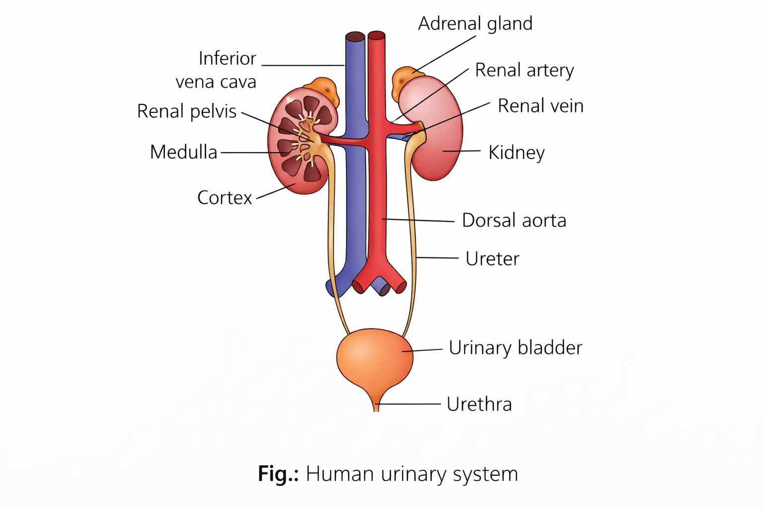
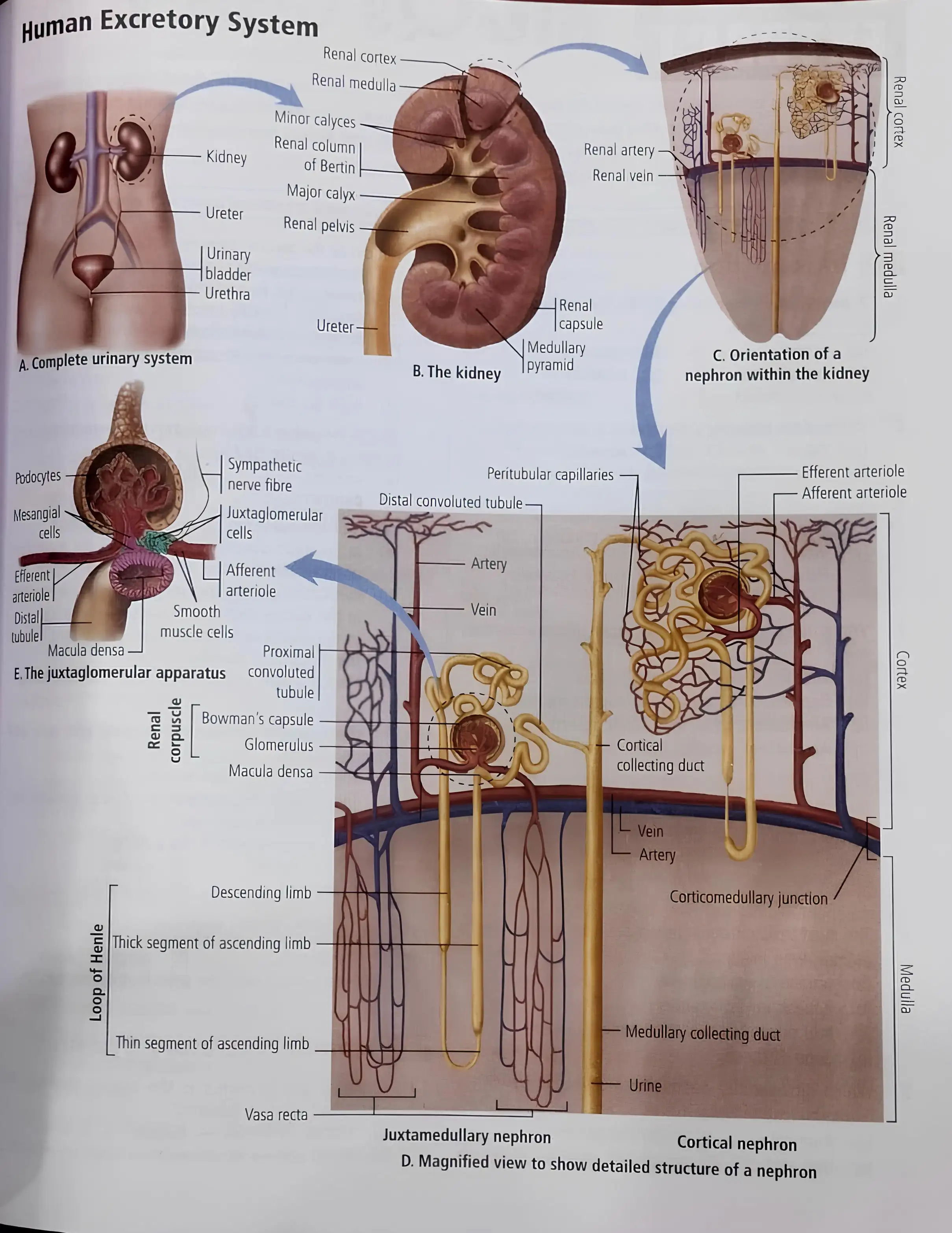
Human excretory system — The system that removes wastes consists of a pair of kidneys, a pair of ureters, the urinary bladder and the urethra.
Kidneys and Nephron Structure
Kidney position and external features
- Shape and position: Kidneys are reddish-brown and bean-shaped, lying near the dorsal inner abdominal wall between the last thoracic and third lumbar vertebrae. The right kidney is slightly lower than the left, below the diaphragm.
- Adult size: Each kidney is about – long, – wide and – thick; average mass is –.
- Hilum: The notch on the inner concave surface through which the ureter, blood vessels and nerves enter or leave.
Internal kidney structure
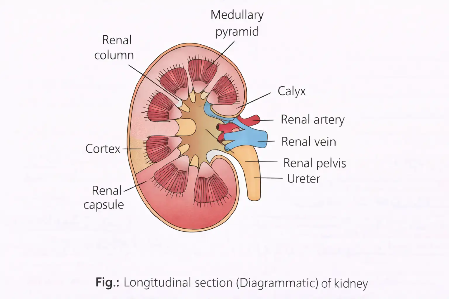
- Layers: A tough capsule surrounds the kidney; inside are an outer cortex and inner medulla.
- Medulla: Divided into conical medullary pyramids that project into the calyces.
- Cortex: Extends between the pyramids as renal columns, also called columns of Bertin.
- Calyces and pelvis: Each human kidney has two or three major calyces; they open into the renal pelvis, which continues as the ureter.
Nephron — The structural and functional unit of the kidney; each kidney contains nearly one million nephrons.
Parts of a nephron
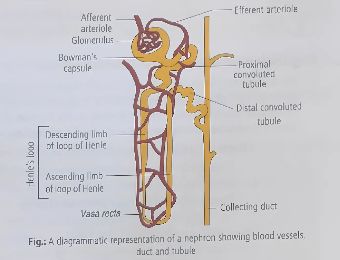
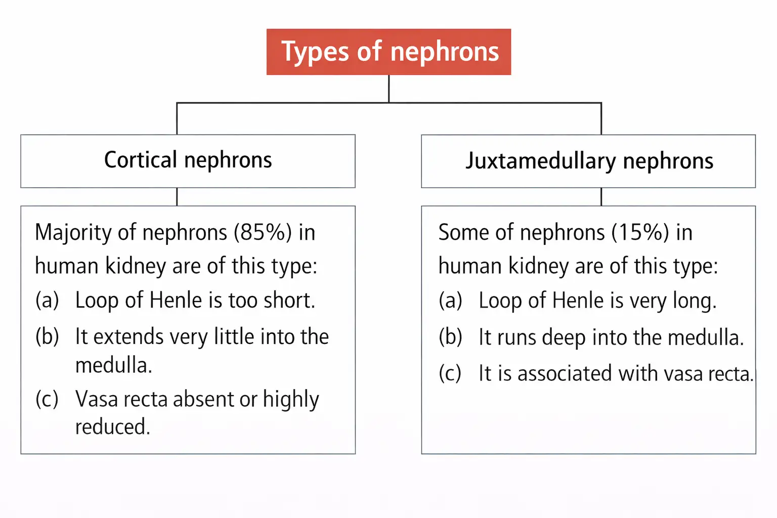
- Two main parts: A nephron consists of a glomerulus and a renal tubule.
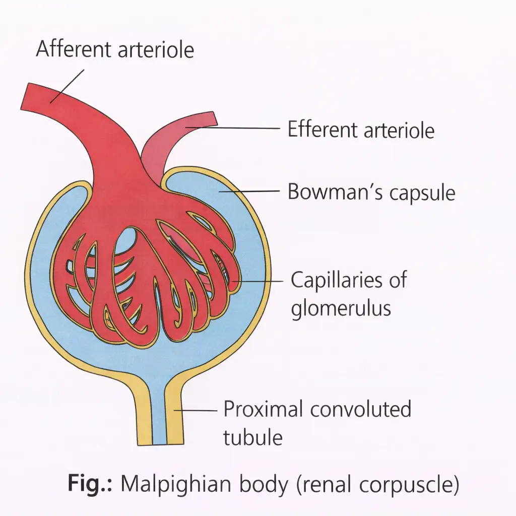
- Glomerulus: A capillary tuft formed from the afferent arteriole, a branch of the renal artery; blood leaves through the efferent arteriole.
- Renal corpuscle: The glomerulus is enclosed by the double-walled, cup-shaped Bowman’s capsule. Together they form the Malpighian body, or renal corpuscle.
- Tubule sequence: Bowman’s capsule → proximal convoluted tubule (PCT) → Henle’s loop → distal convoluted tubule (DCT) → collecting duct.
- Drainage: DCTs join collecting ducts; multiple collecting ducts converge and pass through medullary pyramids to the renal pelvis and calyces.
| Type | Key feature and occurrence |
|---|---|
| Archinephros (archinephric kidney) | Basic ancestral form; found in larvae of some cyclostomes, such as Myxine. |
| Pronephros (pronephric kidney) | Most primitive excretory organ; develops in the embryonic stage of some vertebrates and represents the first kidney-development stage. |
| Mesonephros (mesonephric kidney) | Middle kidney; main excretory organ in aquatic vertebrates and a temporary kidney in higher vertebrates. |
| Metanephros (metanephric kidney) | Most advanced kidney; has a loop of Henle and is functional in reptiles, birds and mammals. |
Nephron regions and blood supply
- Cortex: Contains the Malpighian corpuscle, PCT and DCT.
- Medulla: Contains the loop of Henle, collecting ducts and ducts of Bellini.
- PCT cells: Numerous brush-border microvilli increase the surface area for exchange.
- Descending limb: Long and relatively thin; lined with squamous epithelium, has sparse microvilli and few mitochondria, and is freely permeable to water.
- Ascending limb: Thicker than the descending limb and lined with flattened cuboidal epithelium; it continues into the DCT.
- Peritubular capillaries: The efferent arteriole forms a fine capillary network around renal tubules.
- Vasa recta: A small vessel from this network runs alongside Henle’s loop, forming a -shaped arrangement.
Urine Formation
Main processes
- Glomerular filtration (ultrafiltration)
- Tubular reabsorption
- Tubular secretion
Glomerular Filtration
Glomerular filtration (ultrafiltration) — Pressure-driven filtration of blood plasma from glomerular capillaries into Bowman’s capsule.
Filtration facts
- Blood flow: The kidneys receive about – of blood per minute, roughly one-fifth of the blood pumped by each ventricle per minute.
- Filtration barrier: Filtration occurs through the glomerular capillary endothelium, the basement membrane and the epithelium of Bowman’s capsule.
- Podocytes: Bowman’s capsule epithelial cells form filtration slits (slit pores).
- Filtrate: Water and nearly all plasma constituents pass into the capsule, while plasma proteins are largely retained; hence the process is called ultrafiltration.
- Energy: Filtration is driven by glomerular blood pressure and requires no energy expenditure by kidney cells.
Glomerular filtration rate (GFR) — The volume of filtrate formed by the kidneys per unit time; in a healthy person it is about , or .
Regulation of GFR
Juxtaglomerular apparatus (JGA) — A sensitive region formed where modified cells of the DCT contact the afferent arteriole; it helps regulate GFR.
JGA response to falling GFR
- A fall in GFR activates juxtaglomerular cells.
- Juxtaglomerular cells release renin.
- Renin promotes glomerular blood flow, helping return GFR towards normal.
Effective filtration pressure (EFP) — The net pressure that drives filtration into Bowman’s capsule.
Pressures determining EFP
- GBHP: Glomerular blood hydrostatic pressure; about . It is the chief filtration force and is maintained by the narrower efferent arteriole.
- BCOP: Blood colloidal osmotic pressure due to plasma proteins; about and opposes filtration.
- CHP: Capsular hydrostatic pressure from fluid in Bowman’s capsule; about and opposes filtration.
Subtract the two pressures opposing filtration from glomerular blood hydrostatic pressure.
Renal plasma flow
- RPF: Of the – of blood circulating through both kidneys each minute, about – is plasma; this is the renal plasma flow.
Tubular Reabsorption
Tubular reabsorption — Transfer of useful substances from nephron filtrate back into the blood through peritubular capillaries.
Reabsorption facts
- Extent: About of the filtrate’s materials are reabsorbed, limiting the loss of water, nutrients and ions.
- Mechanisms: Tubular epithelial cells reabsorb substances by active or passive transport.
- Active reabsorption: Glucose, amino acids and are examples.
- Passive reabsorption: Nitrogenous wastes and water in the initial nephron segments are reabsorbed passively.
Tubular Secretion
Tubular secretion — Transfer of substances from peritubular blood into the nephron tubule, using active or passive transport.
Role of tubular secretion
- Secreted substances: Tubular cells secrete , and ammonia into the filtrate.
- Why it matters: Secretion removes substances not filtered from glomerular blood and helps maintain ionic and acid–base balance in body fluids.
- Low-pressure condition: If blood and filtration pressures fall below a critical level, filtration stops and urine formation occurs by tubular secretion alone.
What are the three main processes involved in urine formation?
Define GFR and state its normal value in a healthy person.
How is effective filtration pressure calculated?
Quick recap
- Excretion removes nitrogenous wastes and other metabolic wastes; animals may be ammonotelic, ureotelic or uricotelic.
- The human urinary system contains kidneys, ureters, a urinary bladder and a urethra.
- The nephron comprises a renal corpuscle and a tubule arranged as PCT, Henle’s loop, DCT and collecting duct.
- Urine formation involves filtration, reabsorption and secretion.
- Normal GFR is about ; EFP is about .
- Tubular reabsorption returns about of filtrate materials to blood; secretion supports ionic and acid–base balance.
Function of the Tubules
Proximal Convoluted Tubule (PCT)
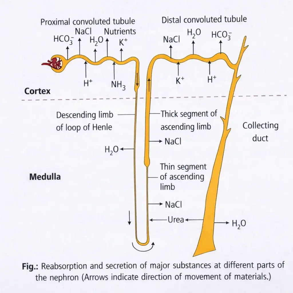
- Reabsorption: The PCT reabsorbs nearly all essential nutrients and about 70–80% of water and electrolytes from the filtrate.
- Secretion: It selectively secretes H⁺, NH₃ and K⁺ and absorbs HCO₃⁻, helping maintain body-fluid pH and ionic balance.
Loop of Henle
- Descending limb: Permeable to water but almost impermeable to electrolytes, so the filtrate becomes concentrated as it descends.
- Ascending limb: Impermeable to water; electrolytes move out, diluting the filtrate and helping maintain the medullary osmolarity.
Distal Convoluted Tubule (DCT)
- Reabsorption: The DCT carries out conditional reabsorption of Na⁺ and water and also reabsorbs HCO₃⁻.
- Secretion: It selectively secretes H⁺, K⁺ and NH₃, helping regulate blood pH and ionic balance.
Collecting Duct
Quick recap
- The PCT has maximum reabsorption; its brush border increases surface area.
- The PCT reabsorbs nearly all essential nutrients and 70–80% of electrolytes and water.
- The descending limb loses water and concentrates filtrate; the ascending limb moves electrolytes out and dilutes it.
- The ascending limb contributes to high medullary interstitial osmolarity and has minimum reabsorption.
- The DCT conditionally reabsorbs and water and selectively secretes ions.
- The collecting duct reabsorbs water, passes some urea into the medulla and secretes and .
Mechanism of Concentration of the Filtrate
Counter-Current Mechanism
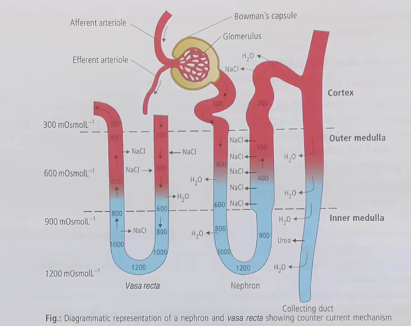
Counter-current mechanism — The opposite flow of filtrate in the two limbs of the loop of Henle, and of blood in the two limbs of the vasa recta, helps establish and maintain the medullary osmotic gradient.
Medullary Osmotic Gradient
- Arrangement: The close proximity of the loop of Henle and vasa recta, together with their counter-current flow, maintains increasing osmolarity towards the inner medulla.
- Gradient: Osmolarity rises from about in the cortex to about in the inner medulla.
- Main contributors: The gradient is produced mainly by NaCl and urea.
Movement of NaCl and Urea
- NaCl moves from the ascending limb of the loop of Henle to the descending limb of the vasa recta.
- NaCl is carried back to the medullary interstitium by the ascending limb of the vasa recta.
- A small amount of urea enters the thin ascending limb of the loop of Henle.
- Urea is transported back to the medullary interstitium by the collecting tubule.
Concentrating the Urine
How the Gradient Concentrates Filtrate
- Counter-current exchange in the loop of Henle and vasa recta maintains high osmolarity in the medullary interstitium.
- The concentrated interstitium allows water to pass out of the collecting tubule.
- Water reabsorption concentrates the filtrate, forming concentrated urine.
Importance of the Loop of Henle
- Function: The loop of Henle helps conserve water.
- Length relationship: In mammals, urine concentration is directly related to the length of the loop of Henle.
- Result: A longer loop allows greater water reabsorption and produces more concentrated urine.
- Capacity: Human kidneys can produce urine nearly four times as concentrated as the initial filtrate.
What maintains the osmotic gradient in the renal medulla?
How does the medullary gradient help concentrate urine?
Quick recap
- Filtrate in the loop of Henle and blood in the vasa recta flow in opposite directions.
- The counter-current mechanism maintains an osmotic gradient from the cortex to the inner medulla.
- NaCl and urea are the main substances contributing to the medullary gradient.
- The gradient enables water reabsorption from the collecting tubule and concentrates urine.
- Longer loops of Henle are associated with greater water reabsorption and more concentrated urine.
- Human kidneys can produce urine nearly four times as concentrated as the initial filtrate.
Regulation of Kidney Function
Antidiuretic Hormone (ADH)
- Release: Fluid loss activates osmoreceptors, prompting the hypothalamus to release ADH (vasopressin) from the neurohypophysis.
- Action: ADH increases water reabsorption in the distal parts of the kidney tubule, reducing urine output; it can also constrict blood vessels and raise blood pressure.
Juxtaglomerular Apparatus (JGA)
Renin–Angiotensin Response
- Reduced glomerular blood flow, blood pressure or GFR
- JG cells release renin
- Renin converts angiotensinogen to angiotensin I, then angiotensin II
- Angiotensin II constricts blood vessels and stimulates aldosterone release
- Aldosterone increases distal tubular reabsorption of and water → blood pressure and GFR rise
Atrial Natriuretic Factor (ANF)
How does ADH help regulate water balance?
How does the renin–angiotensin mechanism raise GFR?
What is the role of ANF in kidney-function regulation?
Quick recap
- Osmoreceptors detect changes in body-fluid volume and ionic concentration.
- ADH promotes distal tubular water reabsorption; its deficiency causes diabetes insipidus.
- Alcohol inhibits ADH release, while caffeine interferes with ADH action and sodium reabsorption.
- Renin initiates the angiotensin pathway when glomerular flow, pressure or GFR falls.
- Angiotensin II and aldosterone raise blood pressure and GFR.
- ANF lowers blood pressure through vasodilation and inhibits renin release.
Micturition
Urine Storage and Micturition
Micturition reflex — The neural mechanisms that bring about the release of urine.
Micturition reflex
- Urine fills bladder → bladder wall stretches.
- Stretch receptors signal the CNS → CNS sends motor messages.
- Bladder smooth muscles contract + urethral sphincter relaxes → urine is expelled.
- Average urine output: An adult excretes about – L of urine per day.
Characteristics of Urine
- Appearance: Urine is a watery, light-yellow fluid; its colour is due to urochrome, formed from the breakdown of haemoglobin.
- Composition: It is slightly acidic, with a pH of about 6, and an adult excretes about 25–30 g of urea per day.
- Odour: Bacteria decompose urea in standing urine to ammonia, giving it a strong smell.
Urine Analysis and Clinical Significance
How does the micturition reflex release urine?
What do glycosuria and ketonuria indicate?
Quick recap
- Micturition is the expulsion of urine; the neural mechanism is the micturition reflex.
- Bladder stretch receptors signal the CNS as urine accumulates.
- CNS motor messages contract bladder smooth muscle and relax the urethral sphincter.
- Adult urine output averages – L daily; urine is slightly acidic, with pH .
- Urochrome gives urine its light-yellow colour, and bacterial breakdown of urea produces ammonia.
- Urine analysis can reveal glycosuria, ketonuria, albuminuria and pyuria.
Role of Other Organs in Excretion
Organs Other Than the Kidneys
- Lungs: They eliminate carbon dioxide and significant quantities of water.
- Skin: Sweat and sebaceous secretions remove small amounts of wastes; saliva can also eliminate small amounts of nitrogenous wastes.
Liver and Bile
- Bile wastes: The liver secretes bilirubin, biliverdin, cholesterol, degraded steroid hormones, vitamins and drugs in bile.
- Elimination: Most of these substances ultimately leave the body with digestive wastes.
Excretion Through the Skin
Sebum — Sebum is an oily secretion from sebaceous glands that protects the skin and carries certain substances out of the body.
Saliva
- Saliva: Small amounts of nitrogenous wastes may also be eliminated through saliva.
Name the substances eliminated by the liver through bile.
What are the contents and main function of sweat?
Quick recap
- The lungs eliminate about 200 mL of carbon dioxide per minute and significant quantities of water.
- The liver secretes waste substances into bile; most leave with digestive wastes.
- Bile carries bilirubin, biliverdin, cholesterol, degraded steroid hormones, vitamins and drugs.
- Sweat contains sodium chloride, small amounts of urea and lactic acid; its main role is body cooling.
- Sebum removes sterols, hydrocarbons and waxes while protecting the skin with an oily covering.
- Small amounts of nitrogenous wastes can also leave through saliva.
Disorders of the Excretory System
Uremia and Hemodialysis
Hemodialysis: Blood Flow
- Blood is drained from a convenient artery and mixed with heparin, an anticoagulant.
- Blood passes through a coiled cellophane tube surrounded by dialysing fluid.
- Nitrogenous wastes diffuse out of the blood into the fluid.
- Cleared blood is returned to the body through a vein after anti-heparin is added.
Kidney Transplantation
- Purpose: Kidney transplantation is the ultimate treatment for acute kidney failure.
- Donor: A compatible donor, preferably a close relative, is selected to minimise the chance of immune rejection.
Renal Calculi
- Definition: Renal calculi are stones or insoluble crystalline salt masses, such as oxalates, formed within the kidney.
- Causes: Dehydration, excess uric acid formation, excess milk intake, metabolic imbalance, infection and hormonal disturbances can contribute to their formation.
Glomerulonephritis
How does hemodialysis remove nitrogenous wastes from blood?
What are renal calculi, and what are their causes?
Quick recap
- Uremia is the harmful accumulation of urea in blood due to kidney malfunction.
- Hemodialysis removes nitrogenous wastes across a porous cellophane membrane.
- Heparin prevents clotting during dialysis; anti-heparin is added before blood returns to the body.
- Kidney transplantation for acute renal failure uses a compatible donor, preferably a close relative.
- Renal calculi are crystallised salt masses formed in the kidney and have several possible causes.
- Glomerulonephritis is inflammation of the kidney glomeruli.
NEET Zoology — ALL CHAPTERS
1
Structural Organisation in Animals
2
Breathing and Exchange of Gases
3
Body Fluids and Circulation
4
Excretory Products and their Elimination
5
Locomotion and Movement
6
Neural Control and Coordination
7
Human Reproduction
8
Reproductive Health
9
Evolution
10
Microbes in Human Welfare
11
Organism and Population
12
Ecosystem
13
Biodiversity and Conservation