NEET UG . NEET Zoology . Zoology . Neural Control and Coordination
Chapter 6: Neural Control and Coordination
Ch 6
NEET Zoology
NEET UG
Neural System
Coordination in the Body
Coordination — The process by which two or more organs interact and complement one another’s functions.
Why coordination is needed
- Homeostasis: Coordination synchronises the functions of organs and organ systems to maintain a stable internal environment.
- Exercise example: Increased muscular activity raises energy and oxygen demand; respiration, heartbeat and blood flow increase to meet it.
- Return to normal: After exercise stops, the activities of nerves, lungs, heart and kidneys gradually return to normal.
| Feature | Neural system | Endocrine system |
|---|---|---|
| Mode of coordination | Point-to-point connections | Chemical integration through hormones |
| Coordination | Quick | Through hormones |
Joint role
- Integration: Neural and endocrine systems jointly coordinate body activities so organs function in a synchronised way.
Neural System
Neuron — A highly specialised cell of the neural system that can detect, receive and transmit different kinds of stimuli.
| Animal group/example | Organisation |
|---|---|
| Hydra (lower invertebrate) | A simple network of neurons |
| Insects | A brain with a number of ganglia and neural tissues |
| Vertebrates | A more developed neural system |
Quick recap
- Coordination enables organs to complement one another and helps maintain homeostasis.
- Exercise increases oxygen demand, leading to increased respiration, heartbeat and blood flow; these activities gradually return to normal after exercise.
- Neural and endocrine systems jointly integrate body activities.
- The neural system uses point-to-point connections for quick coordination; the endocrine system coordinates chemically through hormones.
- Neurons are highly specialised cells that detect, receive and transmit stimuli.
- Neural organisation ranges from a neuron network in Hydra, to a brain with ganglia and neural tissues in insects, to a more developed system in vertebrates.
Human Neural System
Organisation of the Human Neural System
| Division | Components | Main role |
|---|---|---|
| Central neural system (CNS) | Brain and spinal cord | Processes information and controls responses |
| Peripheral neural system (PNS) | All nerves associated with the brain and spinal cord | Connects the CNS with body tissues and organs |
| Fibre type | Direction of impulse | Function |
|---|---|---|
| Afferent | Tissues/organs → CNS | Carries sensory information to the CNS |
| Efferent | CNS → peripheral tissues/organs | Carries regulatory impulses from the CNS |
Functional Divisions of the PNS
| Point | Somatic neural system | Autonomic neural system |
|---|---|---|
| Control | Relays impulses from the CNS to skeletal muscles | Regulates involuntary activities of internal organs |
| Targets | Skeletal muscles | Cardiac muscle, smooth muscle and glands |
| Example | Nerves supplying skeletal muscles | Control of intestinal peristalsis |
Visceral nervous system — The PNS network of nerves, fibres, ganglia and plexuses that carries impulses between the CNS and the viscera.
Peripheral Nerves
| Nerve type | Origin | Number of pairs |
|---|---|---|
| Cranial nerves | Brain | 12 |
| Spinal nerves | Spinal cord | 31 |
Key Features
- Peripheral nerves run outside the brain and spinal cord.
- Myelination: Cranial and spinal nerves are myelinated.
| Cranial nerve | Type |
|---|---|
| Olfactory | Sensory |
| Optic | Sensory |
| Oculomotor | Motor |
| Trochlear | Motor |
| Trigeminal | Mixed |
| Abducens | Motor |
| Facial | Mixed |
| Auditory | Sensory |
| Glossopharyngeal | Mixed |
| Vagus | Mixed |
| Spinal accessory | Motor |
| Hypoglossal | Motor |
| Region | Pairs |
|---|---|
| Cervical | 8 |
| Thoracic | 12 |
| Lumbar | 5 |
| Sacral | 5 |
| Coccygeal | 1 |
- Total spinal nerves: 8 + 12 + 5 + 5 + 1 = 31 pairs.
- Total cranial nerves: 12 pairs.
Autonomic Neural System
Autonomic neural system (ANS) — The self-regulating PNS division that controls involuntary activities of organs, including cardiac and smooth muscles and glands.
| Point | Sympathetic (SNS) | Parasympathetic (PSNS) |
|---|---|---|
| Outflow | Thoracolumbar | Craniosacral |
| Role | Activated during stress; prepares the body for action | Generally has effects antagonistic to the sympathetic division |
| Heart rate | Increases | Opposes sympathetic effects |
| Arteries and blood pressure | Constricts arteries; increases blood pressure | Opposes sympathetic effects |
| Pupil | Dilates | Opposes sympathetic effects |
| Salivation | Decreases | Opposes sympathetic effects |
Functional Organisation
- PNS → somatic neural system → skeletal muscles
- PNS → autonomic neural system → involuntary organs, smooth muscles and glands
- Autonomic neural system → sympathetic division + parasympathetic division
Differentiate between afferent and efferent fibres.
State the number and types of peripheral nerves.
What are the sympathetic and parasympathetic outflows?
Quick recap
- The CNS consists of the brain and spinal cord; the PNS consists of nerves associated with them.
- Afferent impulses travel towards the CNS, while efferent impulses travel away from it.
- The somatic system supplies skeletal muscles; the autonomic system regulates involuntary organs and tissues.
- The PNS has 12 pairs of cranial nerves and 31 pairs of spinal nerves.
- Spinal nerves comprise 8 cervical, 12 thoracic, 5 lumbar, 5 sacral and 1 coccygeal pair.
- The autonomic system includes sympathetic (thoracolumbar) and parasympathetic (craniosacral) divisions with generally antagonistic effects.
Neuron as Structural and Function Unit of Neural System
Neuron: Structure and Function
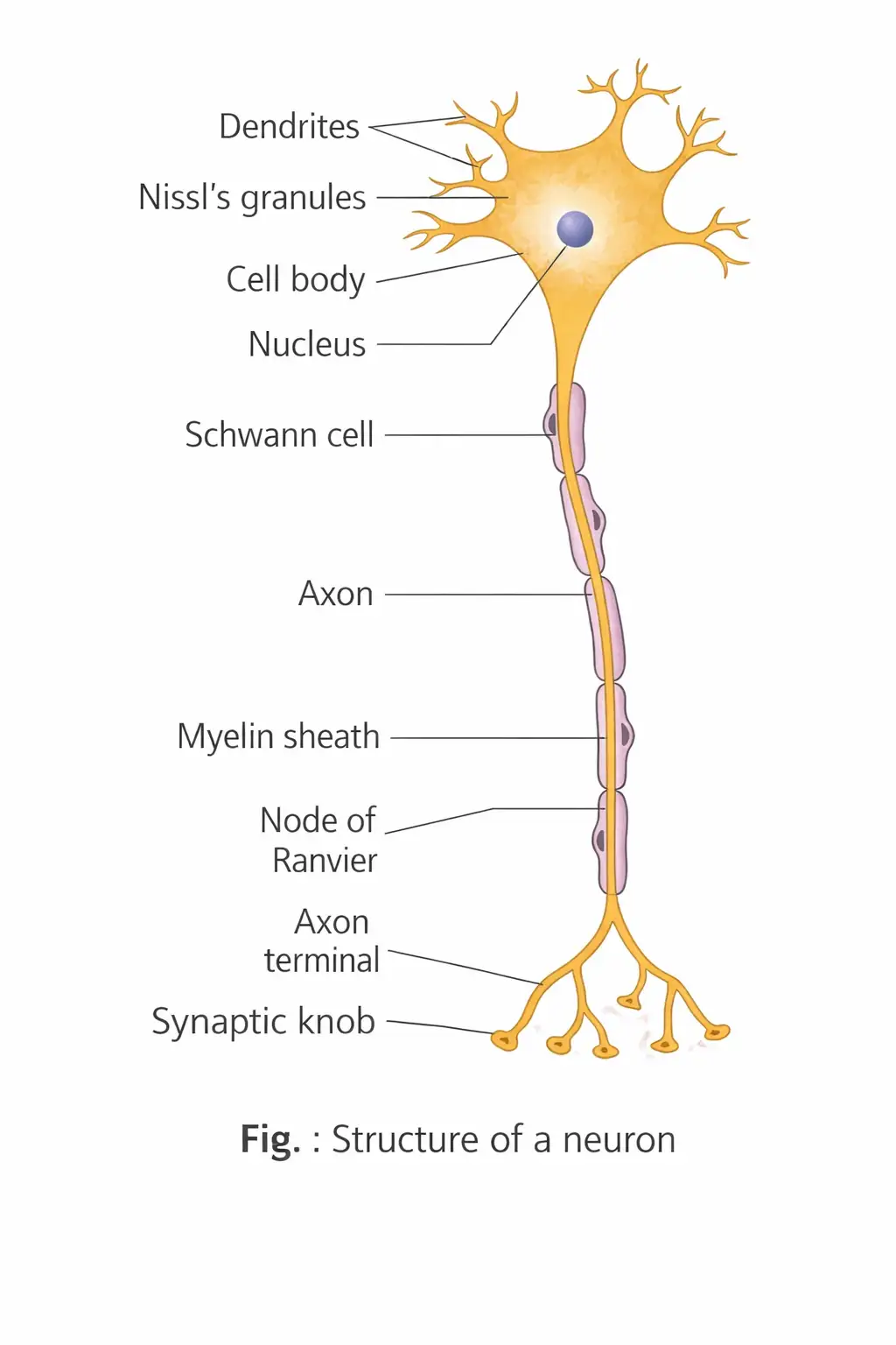
- Neuron: The structural and functional unit of the neural system, specialised to receive and transmit nerve impulses.
- Main parts: A neuron consists of a cell body, dendrites and an axon; dendrites carry impulses towards the cell body, while the axon carries them away.
Types of Neurons
| Type | Structure | Example or location |
|---|---|---|
| Unipolar | Cell body has one axon only. | Usually found in the embryonic stage. |
| Bipolar | One axon and one dendrite. | Retina of the eye. |
| Multipolar | One axon and two or more dendrites. | Cerebral cortex. |
| Pseudounipolar | One axon extends from the cell body and divides into two branches. | Dorsal root ganglion. |
| Non-polar | Processes of the soma are equal; impulses can travel in any direction. | Neurons of coelenterates. |
| Point | Afferent | Efferent |
|---|---|---|
| Function | Carry sensory impulses from receptors to the CNS. | Carry motor impulses from the CNS to effectors such as muscles or glands. |
| Relevant structure | Dendrite terminals may be modified into receptors. | Axon terminals may contact a motor end plate, forming a neuromuscular junction. |
Types of Axons
| Type | Structure | Usual location |
|---|---|---|
| Myelinated | Schwann cells form a myelin sheath around the axon. Gaps between adjacent sheaths are nodes of Ranvier. | Spinal and cranial nerves. |
| Unmyelinated | A Schwann cell encloses the fibre but does not form a myelin sheath. | Autonomic and somatic neural systems. |
Generation and Conduction of a Nerve Impulse
For each cycle, the pump exports three sodium ions and imports two potassium ions.
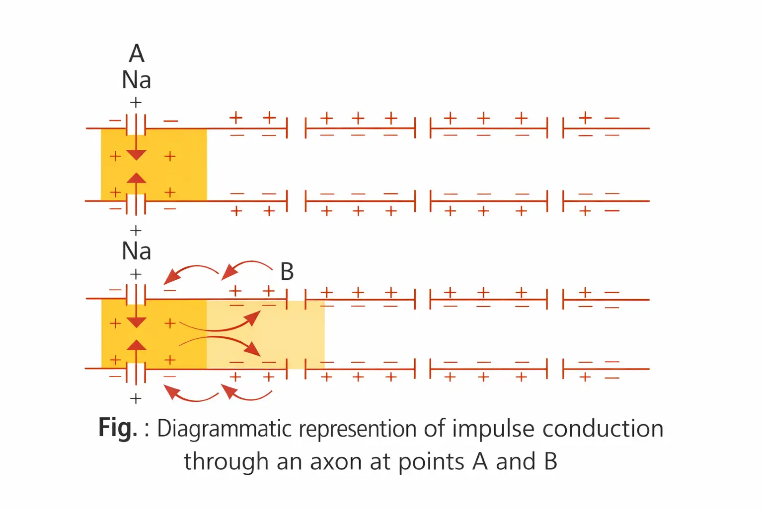
Action potential — The electrical potential difference produced at a stimulated membrane site when its polarity reverses; this is the nerve impulse.
Action Potential and Impulse Conduction
- Stimulus: At the stimulated site, the membrane becomes highly permeable to ; sodium ions rush into the axon.
- Depolarisation: The inner surface becomes positive and the outer surface negative; the action potential reaches about to .
- Local current: Current flows inside the axon from the excited site to the next resting site, and returns along the outer surface.
- Propagation: The next site depolarises; the sequence repeats along the axon, carrying the impulse forward.
- Repolarisation: Increased permeability to allows potassium ions to diffuse out, restoring the resting polarity.
Refractory period — The period of complete inexcitability between depolarisation and repolarisation.
Conduction Features
- Direction: Nerve impulse conduction along an axon is unidirectional.
- Saltatory conduction: In myelinated axons, the action potential jumps from one node of Ranvier to the next, making conduction faster than in unmyelinated axons.
- Unmyelinated fibres: The impulse travels through successive local currents along the membrane.
Transmission of Impulses Across Synapses
| Point | Electrical synapse | Chemical synapse |
|---|---|---|
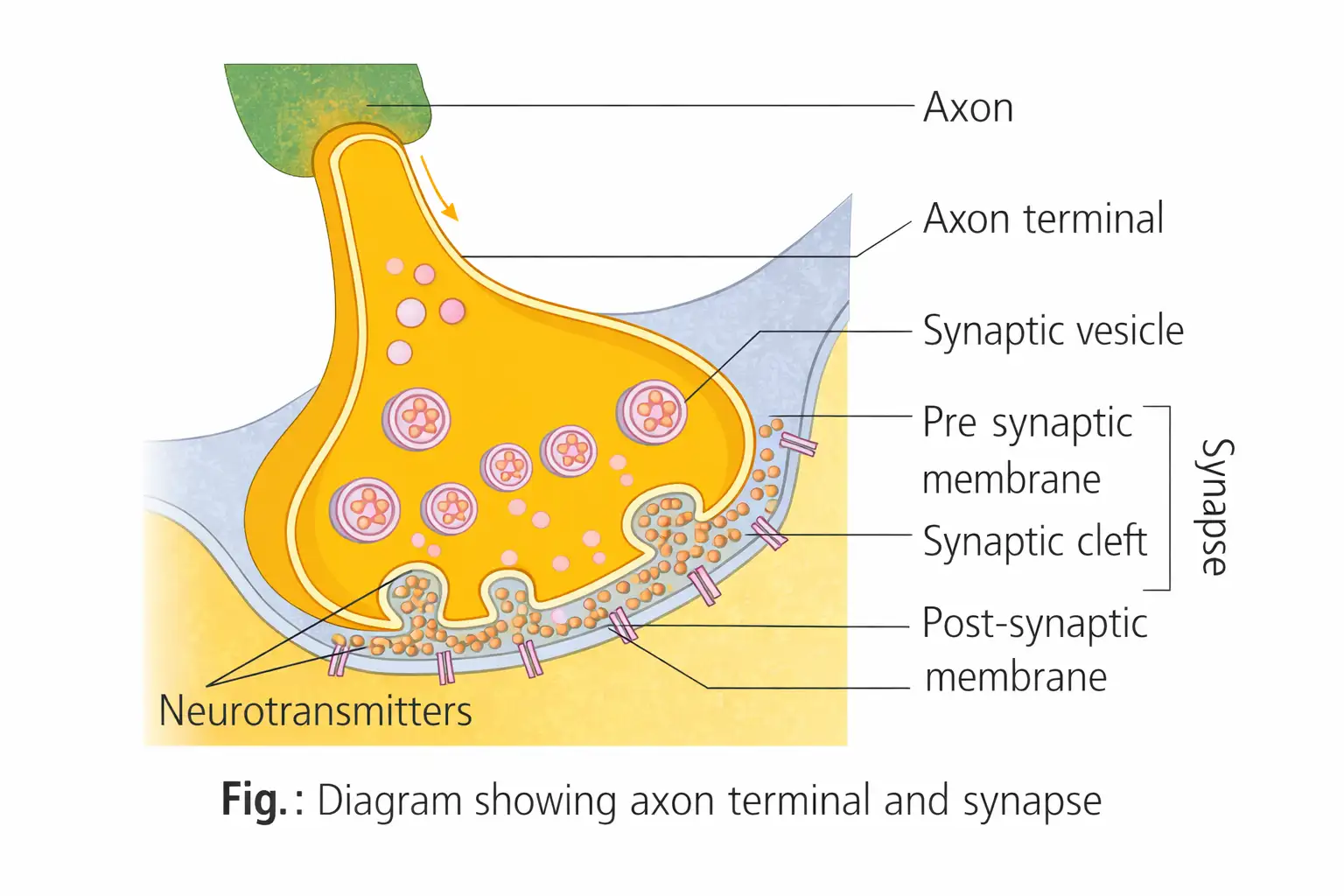
| Connection | Presynaptic and postsynaptic membranes are very close; current passes directly between neurons. | Membranes are separated by a fluid-filled synaptic cleft. |
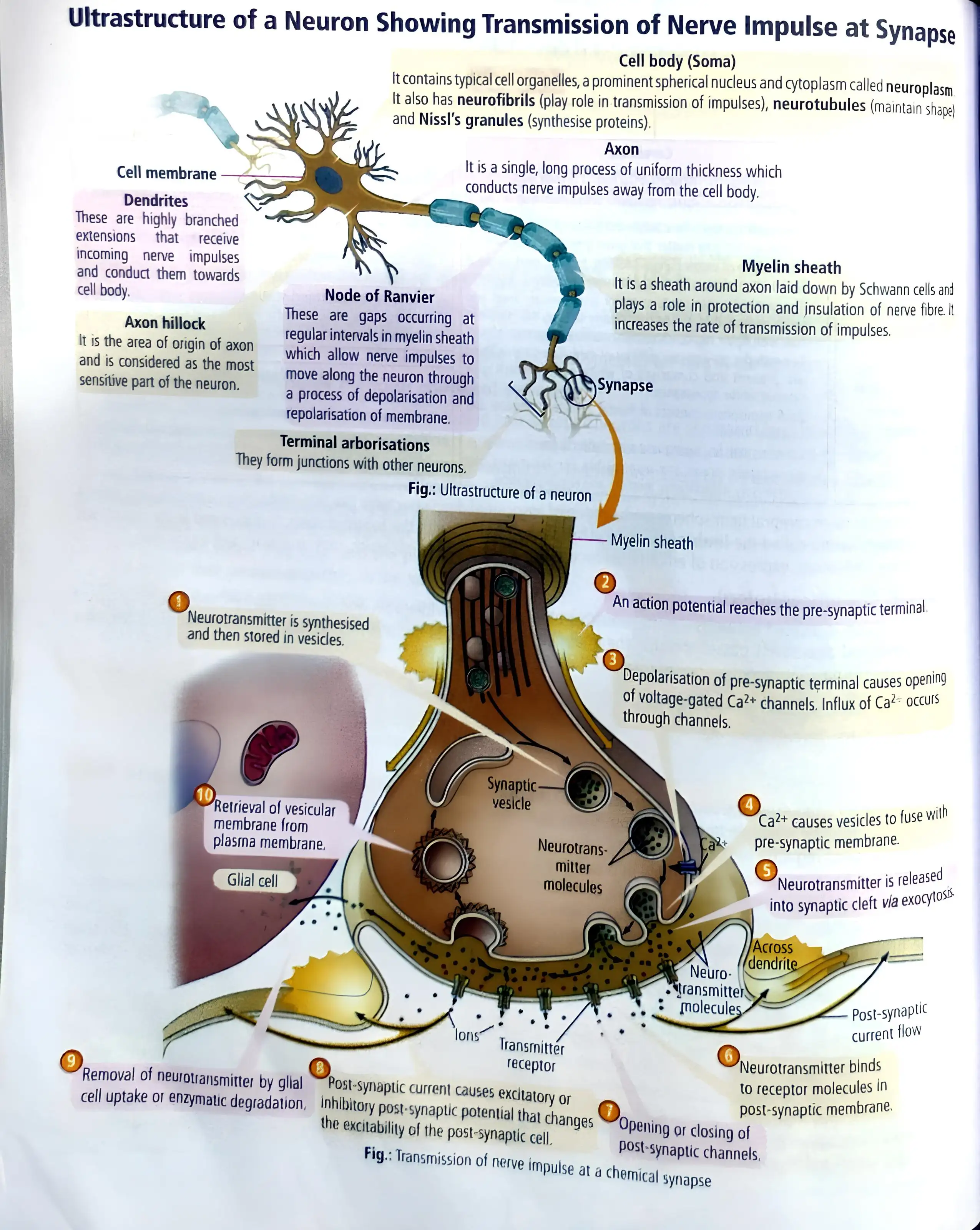
| Transmission | Direct electrical current; similar to conduction along an axon. | Neurotransmitters are released and bind to receptors on the postsynaptic membrane. |
| Speed and occurrence | Faster; rare in the human nervous system. | Slower than electrical transmission. |
Chemical Synaptic Transmission
How is the resting potential maintained?
What is saltatory conduction?
Outline chemical synaptic transmission.
Quick recap
- A neuron has a cell body, dendrites and an axon; dendrites carry impulses towards the cell body and the axon carries them away.
- Neurons are classified by their processes and by their sensory (afferent) or motor (efferent) function.
- Myelin sheaths have gaps called nodes of Ranvier; impulses travel faster by saltatory conduction in myelinated fibres.
- Resting potential is about ; sodium influx causes depolarisation and potassium efflux restores resting polarity.
- The refractory period is the period of complete inexcitability between depolarisation and repolarisation; impulse conduction along an axon is unidirectional.
- Electrical synapses transmit current directly; chemical synapses use neurotransmitters across a synaptic cleft.
Central Neural System
Brain: Role and Protection
Brain — The brain is the body’s central information-processing organ and command-and-control system.
Major functions
- Control: Directs voluntary movement, body balance and vital involuntary functions of organs such as the lungs, heart and kidneys.
- Regulation: Influences thermoregulation, hunger, thirst, circadian rhythms and the activity of several endocrine glands.
- Higher functions: Processes vision, hearing, speech, memory, intelligence, emotions, thoughts and behaviour.
Protection
- Skull: Encloses and protects the brain.
- Cranial meninges: Three protective coverings, arranged from outside to inside as dura mater, arachnoid and pia mater.
- Cerebrospinal fluid (CSF): Clear fluid in the brain’s ventricles and the subarachnoid space around the brain and spinal cord; it cushions the brain against shocks.
Major brain divisions
- Forebrain: Includes the olfactory lobes, cerebrum and diencephalon.
- Midbrain: Lies between the forebrain and hindbrain.
- Hindbrain: Consists of the pons, cerebellum and medulla oblongata.
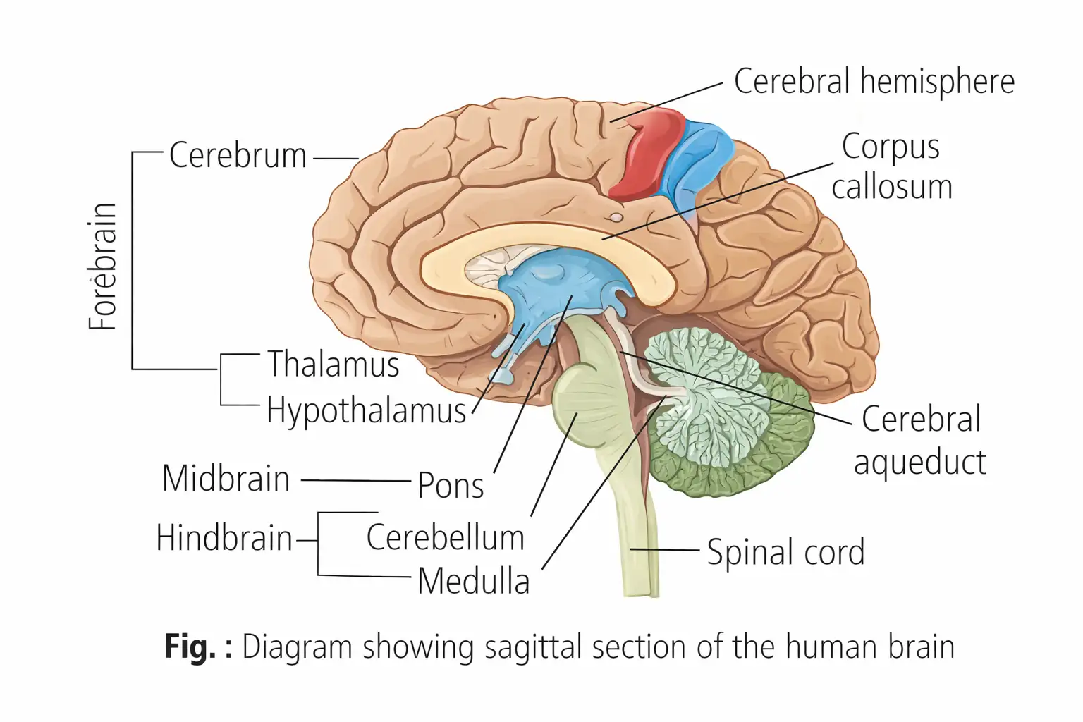
Forebrain (Prosencephalon)
Forebrain structures and functions
- Components: Olfactory lobes, cerebrum and diencephalon.
- Limbic system: The inner parts of the cerebral hemispheres and deep structures such as the amygdala and hippocampus form the limbic lobe/system.
- Role: Along with the hypothalamus, the limbic system helps regulate sexual behaviour, emotional responses (such as excitement, pleasure, rage and fear) and motivation.
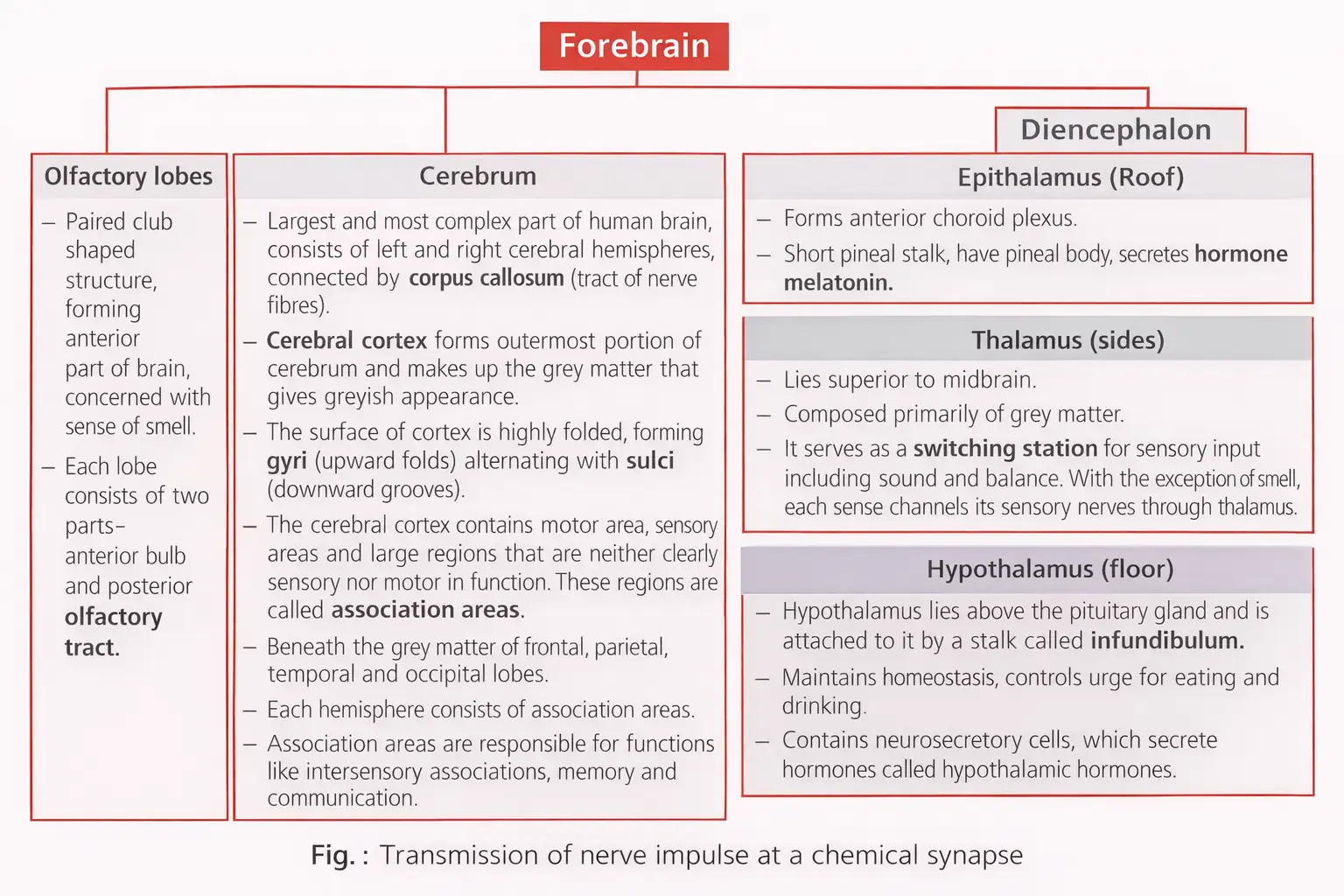
Midbrain (Mesencephalon)
Location and key structures
- Location: Lies between the thalamus/hypothalamus of the forebrain and the pons of the hindbrain.
- Cerebral aqueduct: A canal that passes through the midbrain.
- Corpora quadrigemina: Four dorsal lobes, comprising the superior and inferior colliculi.
- Superior colliculi: Associated with sight; inferior colliculi: associated with hearing.
- Crura cerebri: Relay impulses between the cerebrum, cerebellum, pons and medulla.
Hindbrain
| Part | Key features and functions |
|---|---|
| Pons | Below the midbrain; fibre tracts connect different brain regions. Its pneumotaxic centre regulates breathing. |
| Cerebellum | Its highly convoluted surface provides space for many neurons. Coordinates movement, posture and balance. |
| Medulla oblongata | Connects with the spinal cord; contains centres controlling respiration, cardiovascular reflexes and gastric secretions. |
Brain Stem
Structure and role
- Components: Midbrain, pons and medulla oblongata; it connects the brain to the spinal cord.
- Functions: Supports life-sustaining functions, coordinates and integrates incoming information, and provides a route between the brain and spinal cord.
- Cranial nerves: Serves as the entry or exit point for 10 of the 12 pairs of cranial nerves.
Spinal Cord
Structure and functions
- Extent and size: Continues from the medulla oblongata to the level of the second lumbar vertebra; it is about 42–45 cm long and weighs about 35 g.
- Functions: Conducts impulses to and from the brain, controls most reflex activities, and communicates between the brain and spinal nerves.
- Tissue arrangement: Grey matter lies internally and white matter externally.
- Cauda equina: Spinal nerves occupying the vertebral canal below the second lumbar vertebra.
- Terminal structures: The cord ends as the conus medullaris; the filum terminale arises from it and anchors the cord within the vertebral column.
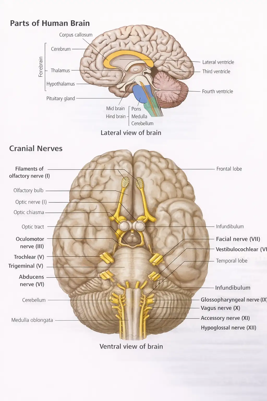
Do not confuse the brain stem with the hindbrain: the hindbrain comprises the pons, cerebellum and medulla, while the brain stem comprises the midbrain, pons and medulla.
Name the three parts of the brain stem and state one role.
Describe the spinal cord’s tissue arrangement and give its main functions.
Quick recap
- The brain processes information and controls voluntary, involuntary and higher functions; the skull, meninges and CSF protect it.
- The forebrain includes the olfactory lobes, cerebrum and diencephalon; the limbic system and hypothalamus regulate emotion, motivation and sexual behaviour.
- The midbrain contains the cerebral aqueduct and corpora quadrigemina; superior colliculi relate to sight and inferior colliculi to hearing.
- The hindbrain comprises the pons, cerebellum and medulla, involved in breathing regulation, coordination and vital reflexes.
- The brain stem is the midbrain, pons and medulla; it connects to the spinal cord and is associated with 10 of 12 cranial nerve pairs.
- The spinal cord extends to the second lumbar vertebra, conducts impulses and reflexes, and has internal grey matter and external white matter.
NEET Zoology — ALL CHAPTERS
1
Structural Organisation in Animals
2
Breathing and Exchange of Gases
3
Body Fluids and Circulation
4
Excretory Products and their Elimination
5
Locomotion and Movement
6
Neural Control and Coordination
7
Human Reproduction
8
Reproductive Health
9
Evolution
10
Microbes in Human Welfare
11
Organism and Population
12
Ecosystem
13
Biodiversity and Conservation