NEET UG . NEET Botany . Botany . Anatomy of Flowering Plants
Chapter 4: Anatomy of Flowering Plants
Ch 4
NEET Botany
NEET UG
The Tissues
Plant Anatomy and Tissues
Meristematic Tissues
- Definition: Meristematic tissue consists of immature cells that actively divide; plant growth is largely restricted to meristems.
- Shape and walls: Cells are generally isodiametric or polygonal, with thin, flexible cellulosic walls.
- Arrangement: Cells are compact, with no intercellular spaces.
- Cell contents: Dense cytoplasm and a prominent nucleus are present; vacuoles are absent or small.
- Term: C. Nägeli coined “meristem” in 1858.
| Type | Origin and role | Examples / outcome |
|---|---|---|
| Promeristem | Embryonic or primordial meristem; appears at the beginning of plant development. | Forms the plant embryo. |
| Primary meristem | Develops from promeristem; appears early and forms the primary plant body. | Apical meristems and intrafascicular cambium; produces primary permanent tissues by differentiation. |
| Secondary meristem | Develops from primary permanent tissue by dedifferentiation; forms the secondary plant body. | Vascular cambium of root, interfascicular cambium, cork cambium and wound cambium; produces secondary permanent tissues by redifferentiation. |
Meristem Classification by Position
- Apical meristem: At root and shoot tips; produces primary tissues and contributes to lengthwise growth.
- Intercalary meristem: Between mature tissues, especially in grasses; helps regenerate parts removed by grazing.
- Lateral meristem: Cylindrical meristems in mature regions of roots and shoots; forms secondary tissues and causes secondary growth.
- Examples of lateral meristems: Fascicular vascular cambium, interfascicular cambium and cork cambium.
- Primary meristems: Apical and intercalary meristems appear early and contribute to the primary plant body.
Apical Meristems and Buds
- Root apical meristem: Occupies the root tip and is protected by the root cap.
- Shoot apical meristem: Occupies the distal end of the stem axis; it produces leaves and helps elongate the stem.
- Axillary bud: Cells left behind by the shoot apical meristem form buds in leaf axils; these can develop into branches or flowers.
- Shoot apex: In longitudinal section, it is conical or dome-shaped and is protected by young leaves.
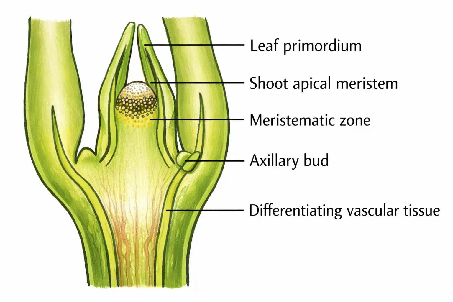
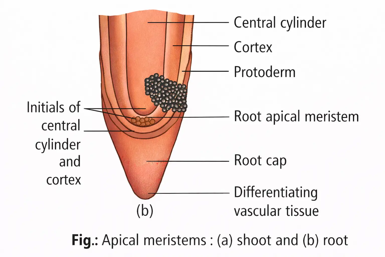
Root Apex and Quiescent Centre
- Root apex: It is subterminal because the root cap protects it, and it bears no lateral appendages.
- Quiescent centre: A group of most distal root cells with a low mitotic rate, often shaped like a hemisphere or disc.
- Cell features: Quiescent-centre cells have light cytoplasm, few mitochondria, less endoplasmic reticulum, and small dictyosomes, nuclei and nucleoli.
- Term: Clowes introduced “quiescent centre” in 1961.
Important distinction: Intrafascicular cambium is the only primary meristem responsible for secondary growth.
Permanent Tissues
| Tissue | Cell features | Location / function |
|---|---|---|
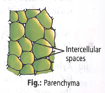
| Parenchyma | Living, usually isodiametric cells; thin cellulosic walls; may be closely packed or have small spaces. | Major component of plant organs; photosynthesis, storage and secretion. |
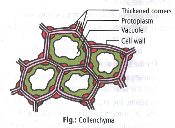
| Collenchyma | Living cells with uneven wall thickening, especially at corners; thickening contains cellulose, hemicellulose and pectin. | Usually in layers or patches below the epidermis of dicots; supports growing parts. |
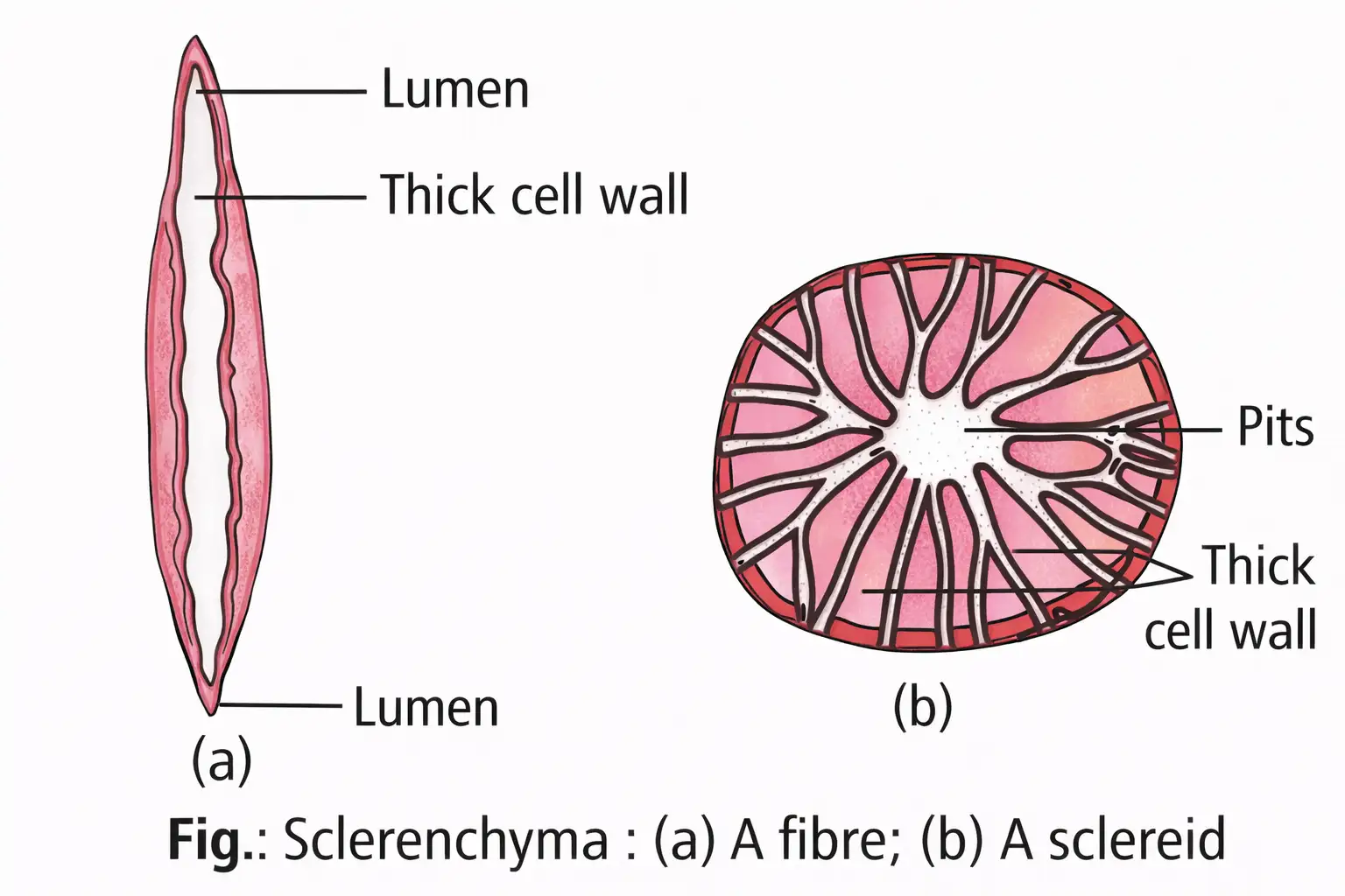
| Sclerenchyma | Usually dead cells with thick, lignified walls and few or many pits. | Provides mechanical support; includes fibres and sclereids. |
Parenchyma and Special Forms
- Cell shapes: Parenchyma cells may be spherical, oval, round, polygonal or elongated.
- Chlorenchyma: Parenchyma with chloroplasts, especially in leaf mesophyll; performs photosynthesis.
- Aerenchyma: Parenchyma with large intercellular spaces in aquatic plants; provides buoyancy to hydrophytes.
Collenchyma
- Location: Commonly forms layers or patches below the epidermis in dicotyledonous plants.
- Structure: Cells may be oval, spherical or polygonal; intercellular spaces are absent and corners are thickened with cellulose, hemicellulose and pectin.
- Function: Provides mechanical support to growing regions, such as young stems and leaf petioles.
- Additional role: Cells containing chloroplasts can assimilate food.
| Point | Fibres | Sclereids |
|---|---|---|
| Shape | Elongated, longest cells in the plant body, with tapering ends. | Spherical, oval or cylindrical. |
| Walls and lumen | Thick walls with numerous simple or bordered pits; lumen present. | Dead cells with heavily lignified, thick walls; lumen very narrow or obliterated. |
| Examples | Coconut coir. | Fruit walls of nuts; pulp of guava, pear and sapota; drupe endocarp; legume seed coats and tea leaves. |
Complex Tissues
- Complex tissues: Made up of more than one type of cell that work together as a functional unit.
- Vascular tissues: Complex tissues are heterogeneous and form the plant’s conducting system.
Xylem
| Element | Structure | Key role / distribution |
|---|---|---|
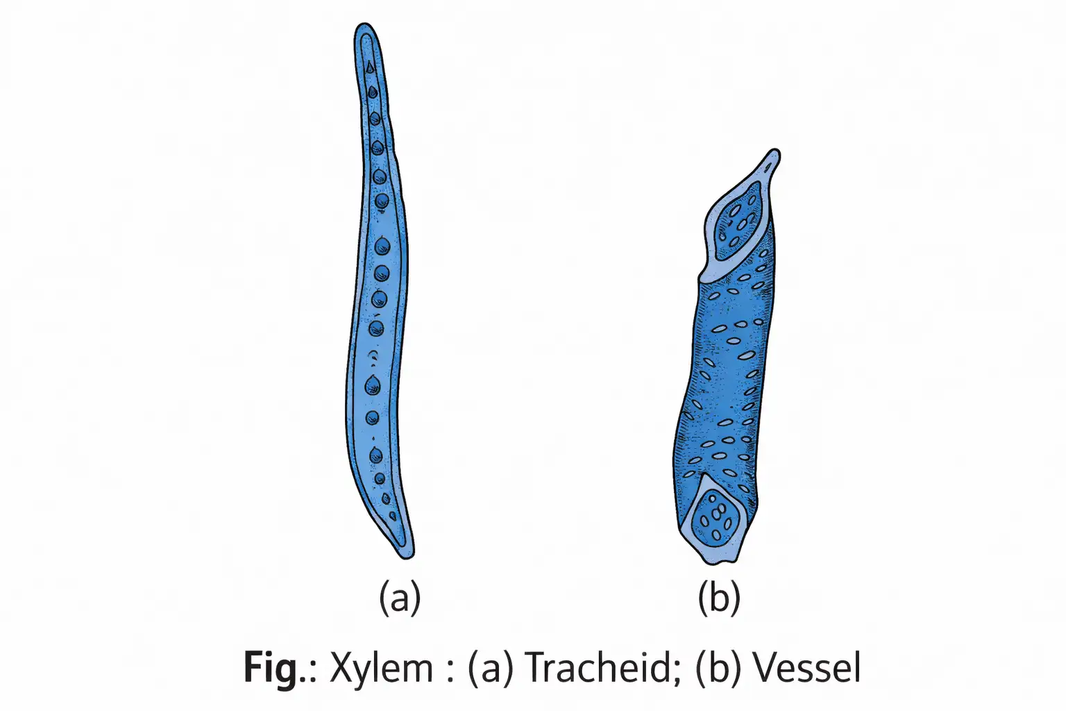
| Tracheids | Elongated, tapering cells with thick lignified walls; dead and without protoplasm. | Primitive conducting elements, especially in pteridophytes and gymnosperms; also conduct water in flowering plants. |
| Vessels | Long cylindrical tubes made of vessel members; lignified walls, large cavity and perforated end walls; dead at maturity. | Advanced conducting elements, characteristic of angiosperms; also occur in Ephedra, Gnetum and Welwitschia. |
| Xylem fibres | Sclerenchyma fibres with highly thickened walls and obliterated central lumen; may be septate or aseptate. | Mechanical support; also called wood fibres. |
| Xylem parenchyma | Living, thin-walled cells with cellulosic walls. | Stores starch, fat, tannins and other materials; ray parenchyma conducts water radially. |
Xylem Wall Thickening
- Tracheid thickening patterns: Annular, spiral, reticulate, scalariform or pitted.
- Protoxylem: Annular and spiral lignin thickening is typical.
- Metaxylem: Reticulate and pitted thickening is typical.
| Type / arrangement | Description |
|---|---|
| Primary xylem | Formed during primary growth; consists of protoxylem (formed first) and metaxylem (formed later). |
| Endarch | In stems, protoxylem lies toward the centre (pith) and metaxylem toward the periphery. |
| Exarch | In roots, protoxylem lies toward the periphery and metaxylem toward the centre. |
| Secondary xylem | Formed during secondary growth; it is not differentiated into protoxylem and metaxylem. |
Phloem
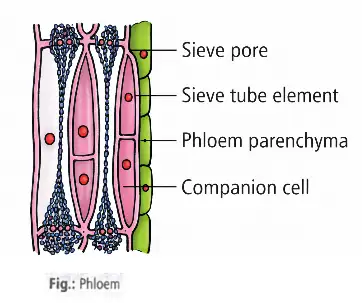
| Element | Structure | Function / key fact |
|---|---|---|
| Sieve tube elements | Long, living tube-like cells arranged longitudinally; end walls have sieve-like perforations forming sieve plates. Mature elements have peripheral cytoplasm and a large vacuole but no nucleus. | Conduct food; their functions are controlled by the companion-cell nucleus. Sieve tubes are described as living syncytes, structures formed by fusion of cells. |
| Companion cells | Specialised parenchymatous cells closely associated with sieve tube elements; connected by pit fields along common longitudinal walls. | Maintain the pressure gradient in sieve tubes. They and their sieve tube element arise from the same meristematic cell and are sister cells. |
| Phloem parenchyma | Elongated, tapering cylindrical cells with dense cytoplasm, a nucleus and cellulosic walls with pits. | Stores food, resins, latex and mucilage; absent in most monocotyledons. |
| Phloem fibres | Long, unbranched cells with thick walls and pointed ends; dead at maturity. | Usually absent from primary phloem but present in secondary phloem; called bast or extraxylary fibres and used commercially. |
Commercial Bast Fibres
- Examples: Jute (*Corchorus capsularis*), sunn hemp (*Crotalaria juncea*), hemp (*Cannabis sativa*) and flax (*Linum usitatissimum*).
| Type | Description |
|---|---|
| Primary phloem | Originates from primary meristem during primary growth; consists of protophloem and metaphloem. |
| Protophloem | First-formed primary phloem; has narrow sieve tubes. |
| Metaphloem | Later-formed primary phloem; has larger sieve tubes. |
| Secondary phloem | Phloem formed during secondary growth. |
Phloem in Pteridophytes and Gymnosperms
- Albuminous cells: Special cells associated with sieve cells in pteridophytes and gymnosperms, in place of companion cells; also called Strasburger cells.
Quick recap
- Meristematic tissues divide; permanent tissues are mature and generally non-dividing.
- Meristems are classified by origin as promeristem, primary and secondary, and by position as apical, intercalary and lateral.
- Intrafascicular cambium is the only primary meristem responsible for secondary growth.
- Parenchyma, collenchyma and sclerenchyma are the three simple tissues; chlorenchyma and aerenchyma are forms of parenchyma.
- Xylem conducts water and minerals; stem xylem is endarch, while root xylem is exarch.
- Phloem transports food; angiosperm phloem includes sieve tubes, companion cells, parenchyma and fibres.
The Tissue System
Classification of Plant Tissue Systems
Tissue system — A group of different tissues that work together as a functional unit to perform a particular plant function.
Types of Tissue Systems
- Classification basis: Their structure and location.
- Three types: Epidermal, ground (fundamental), and vascular (conducting) tissue systems.
Epidermal Tissue System
Epidermal tissue system — The outermost covering of the plant body, made up of epidermal cells, stomata, and epidermal appendages such as trichomes and hairs.
Epidermis
Epidermis — The usually single-layered outermost covering of the primary plant body.
- Cells: Parenchymatous, with little cytoplasm lining the cell wall and a large vacuole.
- Cuticle: A waxy, often thick layer on the outer epidermis that helps prevent water loss.
- Roots: The cuticle is absent.
Stomata
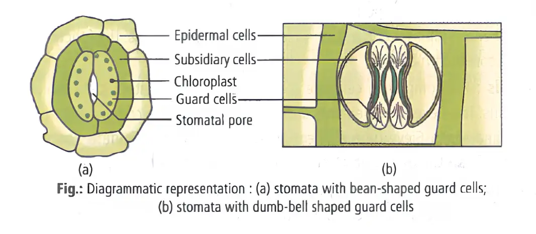
Stomata — Structures in the epidermis of leaves that regulate transpiration and gaseous exchange.
- Guard cells: Each stoma has two guard cells; they are bean-shaped, but dumb-bell-shaped in grasses.
- Wall thickness: The outer walls, away from the pore, are thin; the inner walls facing the pore are highly thickened.
- Function: Guard cells contain chloroplasts and regulate the opening and closing of the stomata.
Stomatal apparatus — The stomatal aperture, guard cells, and surrounding subsidiary cells together form the stomatal apparatus.
Subsidiary Cells
- Location and features: Some epidermal cells near the guard cells become specialised in shape and size; these are subsidiary cells.
Epidermal Appendages
| Feature | Root hairs | Stem trichomes |
|---|---|---|
| Structure | Unicellular elongations of epidermal cells | Usually multicellular epidermal hairs |
| Forms | — | May be branched or unbranched, soft or stiff, and sometimes secretory |
| Role | Absorb water and minerals from the soil | Help prevent water loss through transpiration |
Ground Tissue System
Ground tissue system — The largest tissue system, comprising all tissues except the epidermis and vascular bundles.
- Parts: Hypodermis, cortex, endodermis, pericycle, medullary rays, and pith.
- Tissues: Contains the simple tissues parenchyma, collenchyma, and sclerenchyma.
- In leaves: The ground tissue consists of thin-walled, chloroplast-containing cells called mesophyll.
Vascular Tissue System
Vascular tissue system — The conducting system of xylem and phloem that extends continuously from root to leaf in vascular bundles.
- Vascular bundle: Xylem and phloem together form a vascular bundle.
- Tissue type: Xylem and phloem are complex tissues.
| Feature | Open | Closed |
|---|---|---|
| Cambium | Present between phloem and xylem | Absent |
| Occurrence stated | Dicotyledonous stems | Monocotyledons |
| Secondary tissues | Can form secondary xylem and phloem | Do not form secondary tissues |
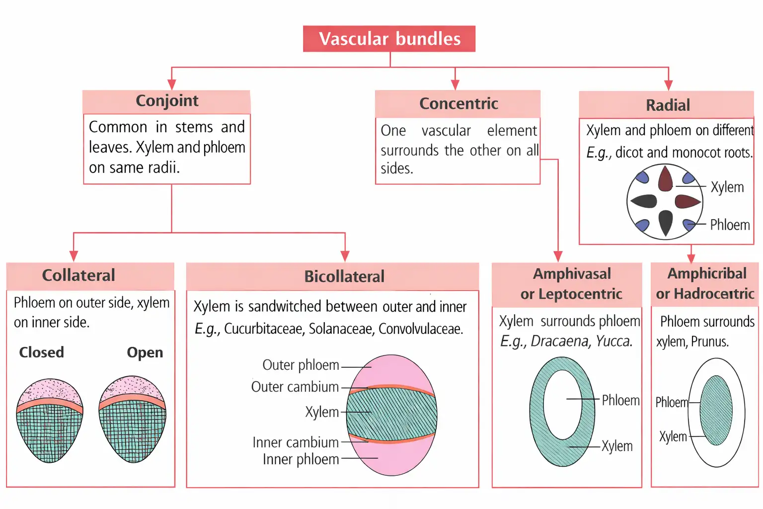
Vascular Bundle Types by Xylem–Phloem Arrangement
- Radial: A vascular bundle type classified by the arrangement of xylem and phloem.
- Conjoint: A vascular bundle type classified by the arrangement of xylem and phloem.
- Concentric: A vascular bundle type classified by the arrangement of xylem and phloem.
Quick recap
- Plant tissue systems are classified as epidermal, ground, or vascular by structure and location.
- The epidermis is usually a single outer layer; its cuticle reduces water loss and is absent in roots.
- Leaf stomata regulate transpiration and gaseous exchange; guard cells control their opening and closing.
- Root hairs absorb water and minerals; stem trichomes may help limit transpiration.
- Ground tissue includes hypodermis, cortex, endodermis, pericycle, medullary rays, and pith; leaf ground tissue is mesophyll.
- Xylem and phloem form vascular bundles; cambium is present in open bundles and absent in closed bundles.
- Radial, conjoint, and concentric bundles are classified by the arrangement of xylem and phloem.
Anatomy of Dicotyledonous and Monocotyledonous Plants
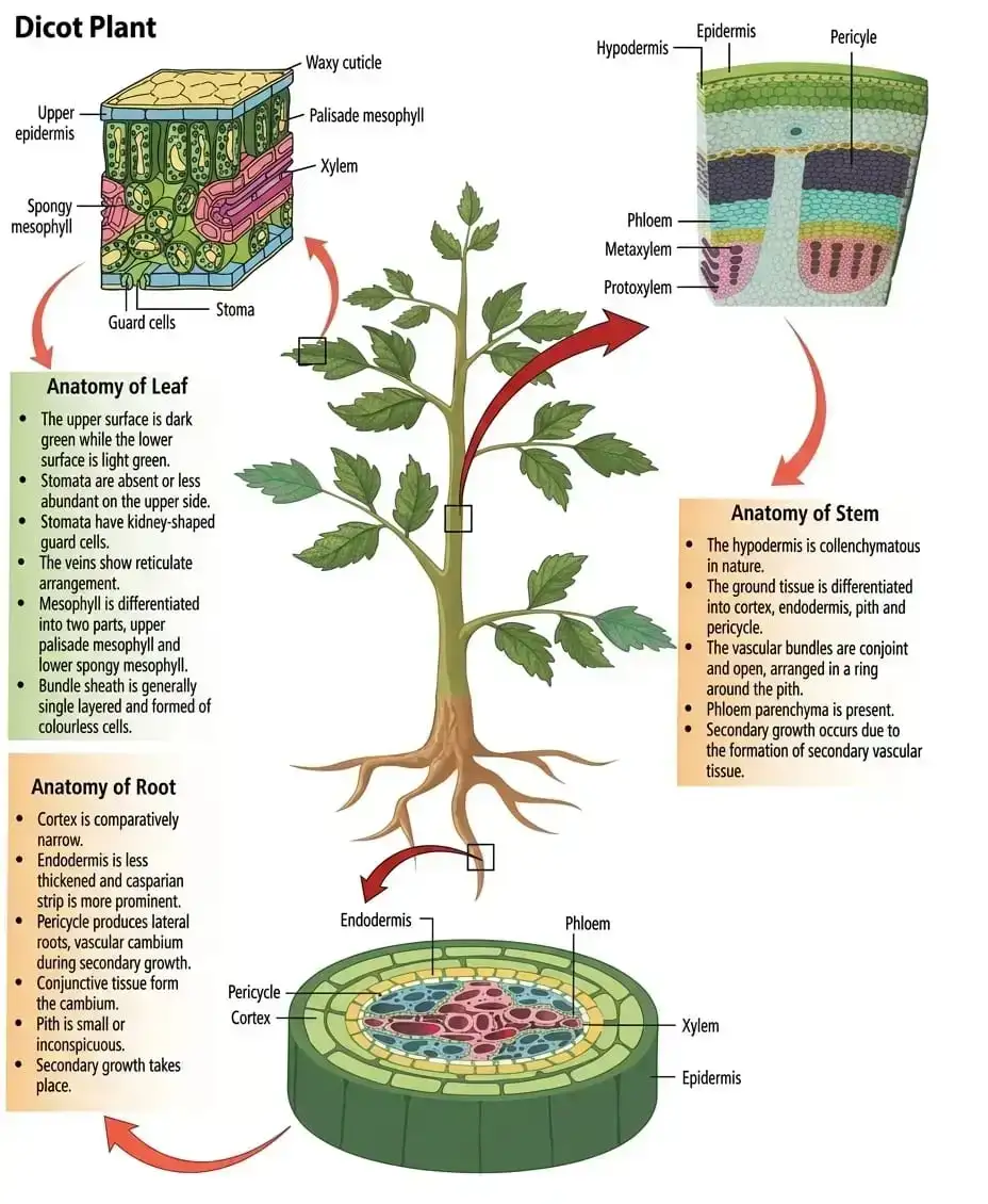
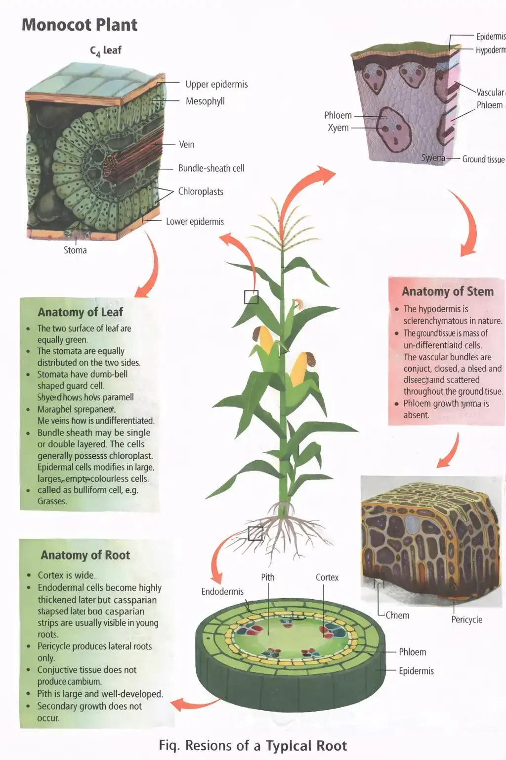
Plant anatomy in transverse section
- Transverse section: A section of a mature root, stem or leaf reveals the arrangement of its tissue layers.
- Anatomical patterns: Dicot and monocot organs differ in characteristic tissue arrangements and vascular-bundle patterns.
Dicotyledonous Root
Tissues and vascular arrangement
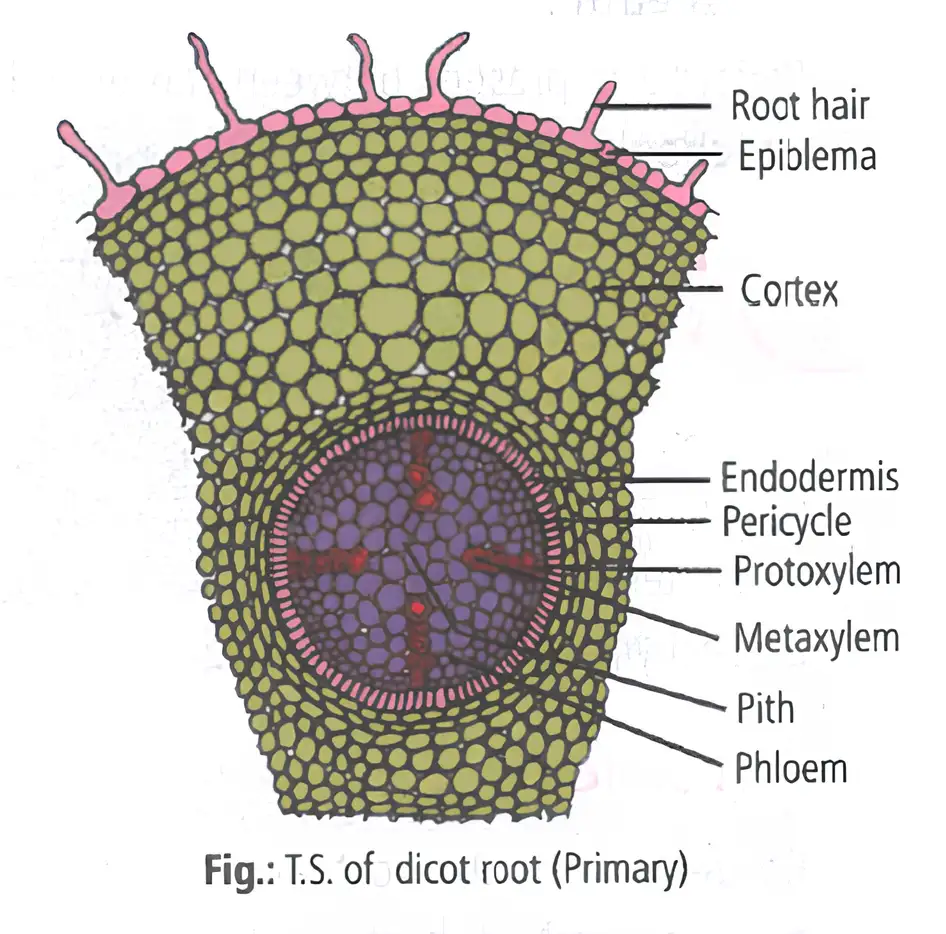
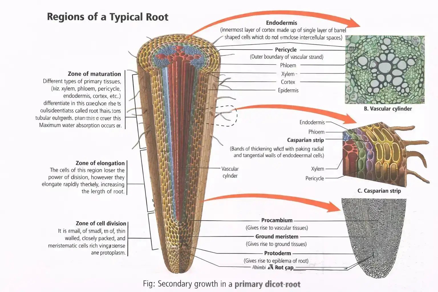
- Epidermis (epiblema): Outermost, single layer; many cells form unicellular root hairs. Roots lack a hypodermis.
- Cortex: Several layers of thin-walled parenchyma with intercellular spaces; its innermost layer is the endodermis.
- Endodermis: Barrel-shaped cells bear Casparian strips on their radial and tangential walls.
- Pericycle: A few layers of thick-walled parenchymatous cells just inside the endodermis; lateral roots arise from it and are therefore endogenous.
- Vascular bundles: Radial arrangement; xylem and phloem alternate and are equal in number. Xylem is usually diarch to tetrarch and exarch.
- Conjunctive tissue: Parenchyma between the xylem and phloem bundles.
- Stele: All tissues inside the endodermis—pericycle, vascular bundles and pith. Its central pith consists of parenchyma.
Monocotyledonous Root
Key features
- Basic plan: Has epidermis, cortex, endodermis, pericycle, vascular bundles and pith, as in a dicot root.
- Xylem: Usually more than six bundles; this condition is called polyarch.
- Pith: Large and well developed.
- Secondary growth: Absent in monocot roots.
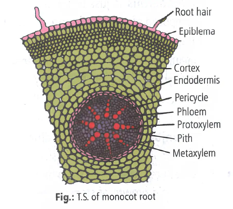
| Feature | Dicot root | Monocot root |
|---|---|---|
| Xylem bundles | Usually 2–4; diarch to tetrarch | Usually more than 6; polyarch |
| Pith | Central parenchymatous pith | Large and well developed |
| Secondary growth | May occur | Absent |
Regions and Special Cells of Roots
Exodermis and passage cells
- Exodermis: In older monocot roots, outer cortical layers become thick-walled and suberised, forming a protective layer.
- Passage cells: Endodermal cells opposite protoxylem groups remain unthickened; they allow fluid movement between the cortex and inner tissues.
Dicotyledonous Stem
Outer tissues and cortex
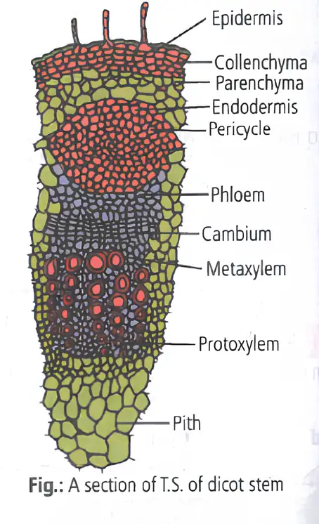
- Epidermis: Outermost, uniseriate layer covered by a cuticle; epidermal cells may form multicellular hairs called trichomes.
- Cortex: Tissue between epidermis and pericycle, differentiated into hypodermis, general cortex and endodermis.
- Hypodermis: Multilayered collenchyma below the epidermis; its cells often contain chloroplasts.
- General cortex: Parenchymatous cells with abundant intercellular spaces.
- Endodermis: Single layer rich in starch grains, hence called the starch sheath.
- Pericycle: Lies between endodermis and vascular bundles, commonly as semicircular patches of sclerenchyma.
Vascular bundles and pith
- Vascular bundles: Numerous bundles arranged in a ring—a characteristic feature of a dicot stem.
- Bundle structure: Each bundle is conjoint, collateral and open; xylem is endarch.
- Pith: The central region contains rounded parenchyma cells with large intercellular spaces.
- Cucurbita: Pericycle is homogeneous, forming a wavy, entirely sclerenchymatous layer about 4–5 cells thick.
- Sunflower: Pericycle is heterogeneous, with alternating bands of parenchyma and sclerenchyma.
Monocotyledonous Stem
Key features
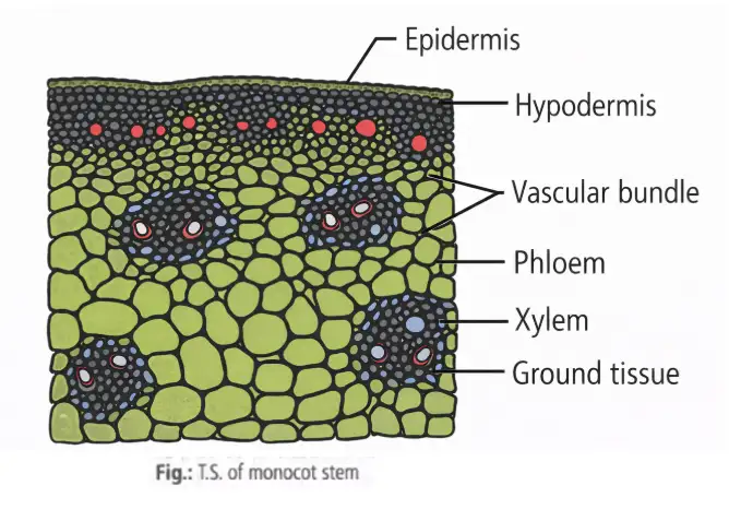
- Epidermis: Uniseriate and covered by a thick cuticle; trichomes are absent.
- Hypodermis: Two to three layers of sclerenchyma that provide mechanical support.
- Ground tissue: Not differentiated into general cortex, endodermis, pericycle, pith or medullary rays.
- Vascular bundles: Numerous, oval and scattered through the ground tissue; each is conjoint, collateral and closed, so secondary growth is absent.
- Bundle sheath: Each vascular bundle is surrounded by sclerenchyma; phloem lies outside and xylem inside.
- Protoxylem cavity (lacuna): Forms when protoxylem vessels and xylem parenchyma dissolve or separate during rapid growth; it stores water.
| Feature | Dicot stem | Monocot stem |
|---|---|---|
| Vascular-bundle arrangement | In a ring | Scattered in undifferentiated ground tissue |
| Bundle type | Conjoint, collateral, open | Conjoint, collateral, closed |
| Xylem position | Endarch | Inside; phloem lies outside |
| Hypodermis | Collenchymatous | Sclerenchymatous |
| Secondary growth | Usually present | Absent |
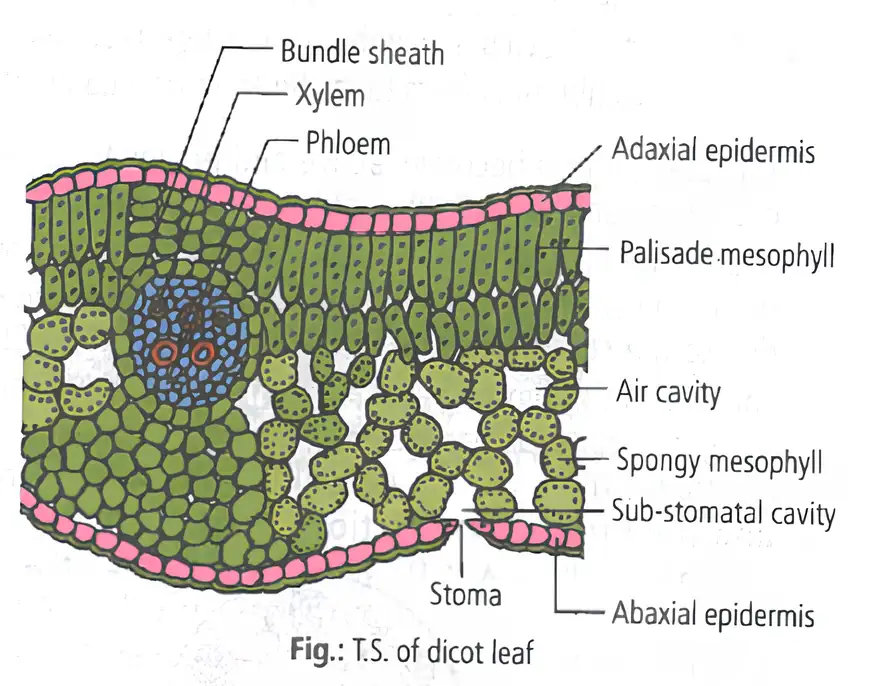
Dorsiventral (Dicotyledonous) Leaf
Leaf tissues and vascular bundles
- Main parts: A dorsiventral (bifacial) leaf has epidermis, mesophyll and a vascular system.
- Epidermis: A single layer covers both surfaces; the upper is adaxial and the lower is abaxial. The lower surface generally has more stomata; both surfaces are covered by a thick cuticle.
- Mesophyll: Chloroplast-containing parenchyma between the epidermises that carries out photosynthesis; it is divided into palisade and spongy tissues.
- Palisade tissue: Elongated parenchyma cells lie vertically and parallel to one another beneath the adaxial epidermis.
- Spongy tissue: Loosely arranged oval or rounded cells occur toward the lower epidermis, with large intercellular spaces and air cavities.
- Bundle sheath: A thin layer of parenchyma surrounds each vascular bundle; larger veins may have sheath extensions toward both leaf surfaces.
- Vascular bundles: Conjoint, collateral and closed; xylem faces the upper epidermis and phloem the lower epidermis. Bundle size varies with vein size.
- Secondary growth: Cambium is absent from leaf vascular bundles, so leaves do not undergo secondary growth.
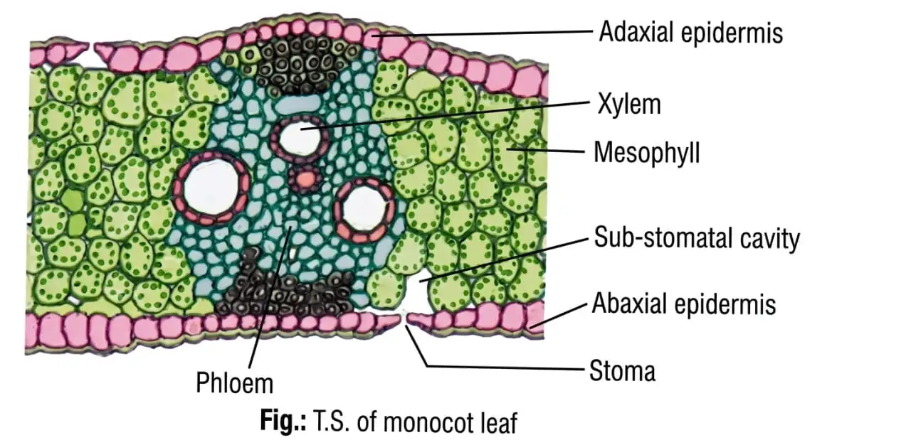
Isobilateral (Monocotyledonous) Leaf
Key features
- Stomata: Present on both epidermal surfaces; this condition is called amphistomatic.
- Mesophyll: Not differentiated into palisade and spongy tissues.
- Bundle-sheath extensions: Sclerenchyma patches above and below large vascular bundles extend to the upper and lower epidermises.
- Bulliform cells: In grasses, large, empty, colourless adaxial epidermal cells along veins; also called motor cells.
- Leaf curling: Turgid bulliform cells expose the leaf surface; when flaccid during water stress, they cause inward curling that reduces water loss.
- Vascular bundles: Parallel venation is reflected in bundles of nearly similar size, except in the main veins.
| Feature | Dorsiventral dicot leaf | Isobilateral monocot leaf |
|---|---|---|
| Stomata | Generally more numerous on lower surface | Present on both surfaces |
| Mesophyll | Differentiated into palisade and spongy tissues | Not differentiated into palisade and spongy tissues |
| Vascular bundles | Vary in size with vein size | Nearly similar in size, except in main veins |
| Special feature | Larger-vein bundle sheath may extend toward both surfaces | Sclerenchyma bundle-sheath extensions reach both surfaces; grasses have bulliform cells |
Secondary Growth
Primary growth — Increase in root and stem length produced by the apical meristem.
Secondary growth — Increase in girth, common in dicot roots and stems, produced by the lateral meristems: vascular cambium and cork cambium.
Secondary Growth in Dicot Stem
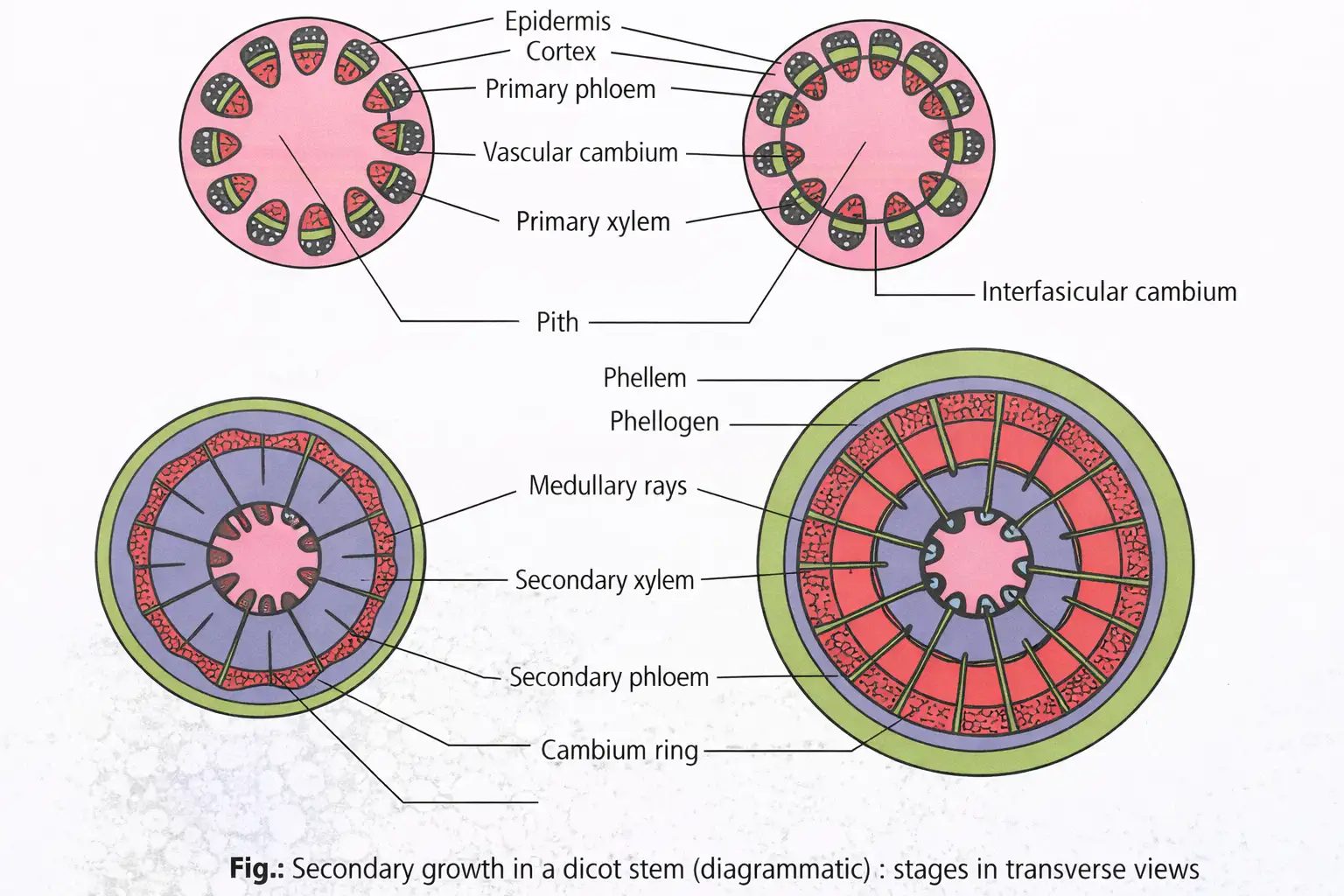
Vascular cambium ring and secondary tissues
- Vascular cambium: Meristematic tissue that produces new xylem and phloem.
- Intrafascicular cambium: In a young stem, a single cambial layer occurs in patches between xylem and phloem within vascular bundles; it is primary in origin.
- Interfascicular cambium: After primary growth, medullary-ray cells beside the intrafascicular cambium become meristematic and form this secondary lateral meristem.
- Cambial ring: Intrafascicular and interfascicular cambia join to form a continuous, single-layered ring. Dicot-stem vascular cambium is partly primary and partly secondary in origin.
- Secondary tissues: The ring cuts cells inward, which mature into secondary xylem, and outward, which mature into secondary phloem.
- Unequal production: Cambium is more active inward, producing more secondary xylem than secondary phloem. The growing xylem forms a compact mass and gradually crushes primary and secondary phloem.
- Primary xylem: Remains more or less intact near the centre.
- Secondary medullary (vascular) rays: Radial bands of parenchyma pass through secondary xylem and phloem, conducting materials in the radial direction.
Formation of secondary vascular tissues
- Intrafascicular cambium joins with interfascicular cambium.
- A continuous vascular cambium ring forms.
- The cambium produces secondary xylem inward and secondary phloem outward.
- Greater inward production accumulates secondary xylem and gradually crushes the phloem.
Spring Wood and Autumn Wood
Annual rings
- Cause: Vascular cambium changes its activity during the year in response to physiological and environmental factors.
- Annual ring: Alternate concentric bands of spring wood and autumn wood; counting rings in a cut stem estimates a tree's age.
- Dendrochronology: Determining a tree's age by counting its annual rings.
| Feature | Spring wood (early wood) | Autumn wood (late wood) |
|---|---|---|
| Cambium activity | Very active | Less active |
| Xylary elements | Many; vessels have wide cavities | Fewer; vessels are narrow |
| Appearance and density | Lighter in colour; lower density | Darker in colour; higher density |
Heartwood and Sapwood
| Feature | Heartwood (duramen) | Sapwood (alburnum) |
|---|---|---|
| Position and colour | Central, older secondary xylem; dark brown | Peripheral secondary xylem; lighter |
| Contents and cells | Dead, highly lignified elements; tannin, resin, oil and aromatic substances accumulate in tracheids and vessels | Peripheral region of secondary xylem |
| Function | Does not conduct water; provides mechanical support | Conducts water and minerals from roots to leaves |
| Properties | Hard, durable and resistant to microorganisms and insects; does not imbibe rainwater | Active in conduction |
Why heartwood does not conduct water
- Organic deposits: Substances gradually fill the cavities of vessels and tracheids.
- Tyloses: Balloon-like ingrowths from adjacent parenchyma cells enter vessel lumens and block conduction.
- Wood quality: Heartwood's durability and resistance to insects and termites make it high-quality wood.
Cork Cambium and Periderm
Formation and products
- Cork cambium (phellogen): A secondary meristem in the extrastelar region; it consists of a few layers of narrow, thin-walled, nearly rectangular cells.
- Division: Phellogen divides periclinally, cutting cells off on both sides.
- Outer product: Outward cells differentiate into cork (phellem); suberin in their walls makes cork impermeable to water.
- Inner product: Inward cells differentiate into parenchymatous secondary cortex (phelloderm).
- Periderm: Phellogen, phellem and phelloderm together form the periderm.
- Commercial cork: Obtained from oak, Quercus suber; it is used to make common bottle cork.
- Sloughing: Pressure from cork-cambium activity causes outer layers to die and peel off.
- Annual replacement: A cork-cambium ring remains active for one year; a new one forms below it each year from the secondary cortex or phelloderm.
Bark — A non-technical term for all tissues outside the vascular cambium, including secondary phloem.
Bark and lenticels
- Bark types: Bark formed early in the season is early or soft bark; bark formed later is late or hard bark.
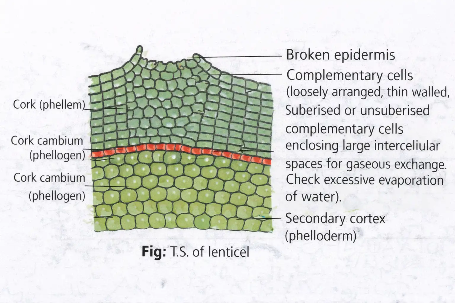
- Lenticels: At some sites, phellogen produces loosely arranged parenchyma outward instead of cork; the cells rupture the epidermis to form a lens-shaped opening.
- Function: Lenticels permit gas exchange between the atmosphere and internal stem tissues; they occur in most woody trees.
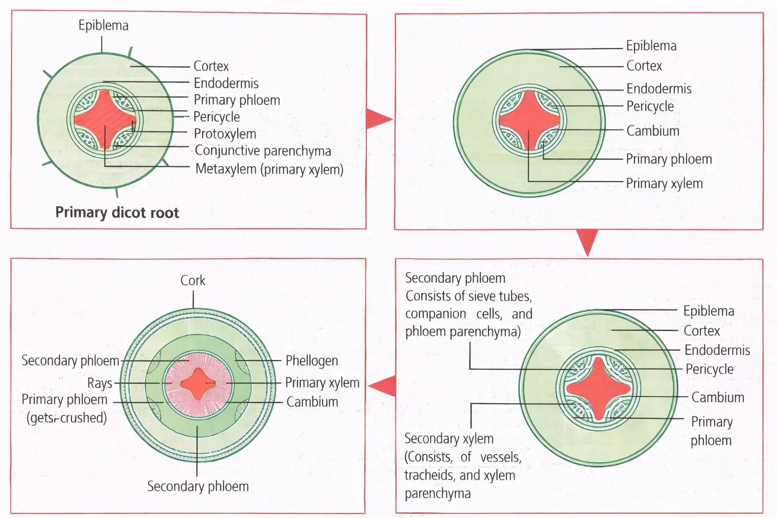
Secondary Growth in Dicot Root
Origin of vascular cambium
- Origin: Dicot-root vascular cambium is entirely secondary in origin.
- Contributors: It arises from tissue just below the phloem bundles and from pericycle portions above the protoxylem.
- Cambial ring: These tissues form a continuous, wavy ring that later becomes circular.
- Further growth: Subsequent secondary-growth events are similar to those in the dicot stem.
How does the vascular cambium ring form in a dicot stem, and what tissues does it produce?
Distinguish heartwood from sapwood by position and function.
What are annual rings, and what is dendrochronology?
Quick recap
- Dicot roots are usually diarch to tetrarch and exarch; monocot roots are polyarch, with a large pith and no secondary growth.
- Dicot stems have ring-arranged open vascular bundles; monocot stems have scattered, closed bundles and undifferentiated ground tissue.
- Dicot leaves have palisade and spongy mesophyll; monocot leaves have undifferentiated mesophyll and often bulliform cells.
- Vascular cambium produces secondary xylem inward and secondary phloem outward; secondary xylem accumulates faster.
- Spring wood is lighter and less dense; autumn wood is darker and denser. Their bands form annual rings.
- Heartwood supports but does not conduct; sapwood conducts water and minerals.
- Cork cambium forms phellem outward and phelloderm inward; lenticels permit gas exchange.
NEET Botany — ALL CHAPTERS
1
The Living World
2
Plant Kingdom
3
Morphology of Flowering Plants
4
Anatomy of Flowering Plants
5
The Unit of Life
6
Photosynthesis in Higher Plants
7
Respiration in Plants
8
Plant Growth and Development
9
Sexual Reproduction in Flowering Plants
10
Principles of Inheritance and Variation
11
Molecular Basis of Inheritance
12
Biotechnology :Principles and Processes
13
Biotechnology and its Applications