NEET UG . NEET Botany . Botany . The Unit of Life
Chapter 5: The Unit of Life
Ch 5
NEET Botany
NEET UG
What is a Cell
Cells and the Unity of Life
Cellular organisation
- Biology is the study of living organisms; their diverse forms share a common cellular organisation.
- Cell theory revealed the unity of life by showing that living organisms are organised into cells.
- Cellular integrity is essential for physiological and behavioural processes to occur.
- Unicellular organisms consist of one cell; multicellular organisms consist of many cells.
- Examples: bacteria are microscopic unicellular organisms; elephants and humans are multicellular.
What Is a Cell?
Cell — The smallest entity capable of independent existence and of carrying out all essential functions of life; it is the basic structural and functional unit of life.
| Scientist | Contribution |
|---|---|
| Robert Hooke | First observed cells in a cork slice. He saw cell walls enclosing spaces left by dead protoplasts. |
| Anton von Leeuwenhoek | First to observe, describe and sketch a free-living cell; observed bacteria, protozoa, spermatozoa and red blood cells. |
| Robert Brown (1831) | Discovered the nucleus in cells of an orchid root. |
| Max Knoll and Ernst Ruska (1931) | Invented the electron microscope. |
Microscopy
- Microscopes and their gradual improvement revealed the structural details of cells.
- Leeuwenhoek is known as the father of microscopy.
Why is the cell called the basic unit of life?
What did Robert Hooke observe in cork?
Quick recap
- Cellular organisation is the common feature uniting living organisms.
- One-celled organisms are unicellular; many-celled organisms are multicellular.
- The cell is the smallest independently existing unit that performs essential life functions.
- Hooke first observed cells in cork; Leeuwenhoek observed free-living cells.
- Brown discovered the nucleus in orchid root cells in 1831.
- Knoll and Ruska invented the electron microscope in 1931.
Cell Theory
Development of Cell Theory
- Development: Cell theory emerged from observations of plant and animal cells by Matthias Schleiden and Theodor Schwann.
- Refinement: Rudolf Virchow later added that new cells arise from pre-existing cells.
Main Statements and Exception
- Cellular composition: All living organisms are composed of cells and cell products.
- Cell origin: All cells arise from pre-existing cells (Omnis cellula-e cellula).
- Exception: Viruses are not cellular and are exceptions to cell theory.
Quick recap
- Schleiden observed that plants are composed of cells (1838).
- Schwann reported a plasma membrane in animal cells (1839).
- Schleiden and Schwann formulated cell theory.
- Virchow stated that new cells arise from pre-existing cells (1855).
- All organisms consist of cells and cell products; viruses are the exception because they lack cellular organisation.
An Overview of Cell
Cell Boundaries and Internal Organisation
Basic organisation
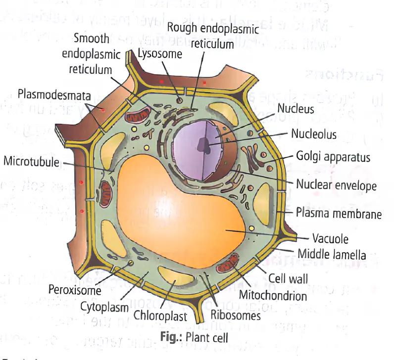
- Plant cell boundary: The cell wall is the outer boundary; the cell membrane lies just inside it.
- Human cell boundary: The cell membrane forms the cell’s outer delimiting structure.
- Nucleus: A membrane-bound structure containing chromosomes, which carry DNA.
- Cytoplasm: A semi-fluid matrix filling the cell; it is the main arena of cellular activities and contains reactions that maintain the living state.
| Feature | Eukaryotic cells | Prokaryotic cells |
|---|---|---|
| Nucleus | Membrane-bound nucleus present | Membrane-bound nucleus absent |
| Membrane-bound organelles | Present | Absent |
| Cytoplasm and ribosomes | Present | Present |
Membrane-Bound Organelles
- Eukaryotic organelles: Besides the nucleus, these include the endoplasmic reticulum (ER), Golgi complex, lysosomes, mitochondria, microbodies and vacuoles.
- Location: These organelles are embedded in the cytoplasm.
Non-Membrane-Bound Organelles
Ribosomes and Centrioles
- Ribosomes: Non-membrane-bound organelles found in both prokaryotic and eukaryotic cells.
- Locations: Ribosomes occur in the cytoplasm, inside mitochondria and chloroplasts, and attached to rough ER.
- Organelle within an organelle: Ribosomes are also found inside mitochondria and chloroplasts.
- Centriole: An additional non-membrane-bound organelle in animal cells that helps in cell division.
Cell Size and Shape
Variation in Cell Size
- Smallest cells: Mycoplasmas, about long.
- Bacteria: About –.
- Largest isolated single cell: The egg of an ostrich.
- Human red blood cells: About in diameter.
- Among the longest cells: Nerve cells.
Variation in Cell Shape
- Possible shapes: Disc-like, polygonal, columnar, cuboid, thread-like or irregular.
- Shape and function: A cell’s shape may vary according to the function it performs.
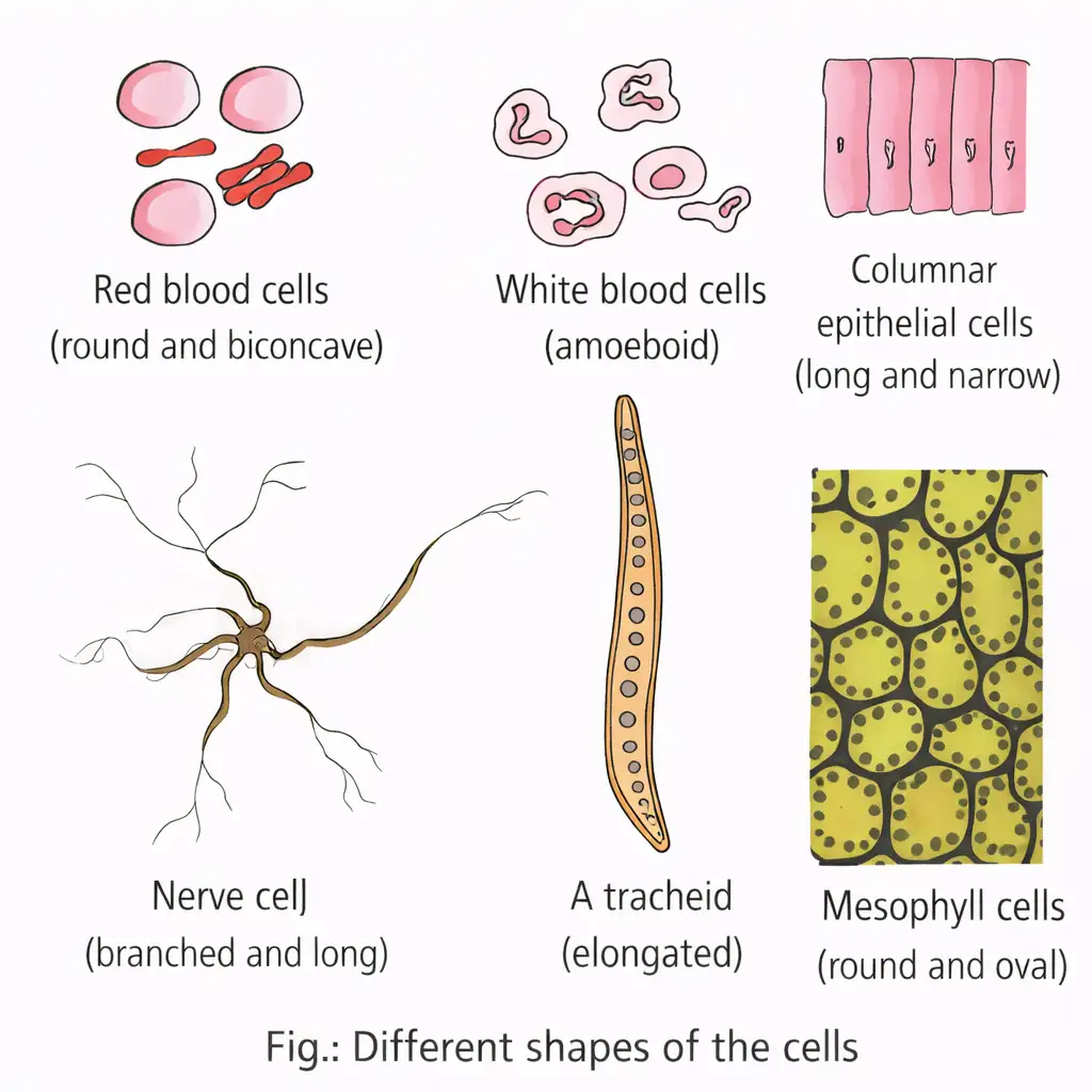
Quick recap
- Plant cells have a cell wall outside the cell membrane; human cells are bounded by the cell membrane.
- Eukaryotic cells have a membrane-bound nucleus and membrane-bound organelles; prokaryotic cells lack both.
- Cytoplasm and ribosomes are present in both prokaryotic and eukaryotic cells.
- Ribosomes occur in the cytoplasm, mitochondria, chloroplasts and on rough ER.
- Cell sizes vary: Mycoplasmas are among the smallest, while the ostrich egg is the largest isolated single cell.
- Cell shape may vary with the function performed.
Prokaryotic Cells
Prokaryotic Cell: Features and Forms of Bacteria.
Prokaryotic cell — A cell without a well-defined, membrane-bound nucleus; examples include bacteria, blue-green algae (cyanobacteria), mycoplasma and PPLO (Pleuro Pneumonia-Like Organisms).
Key Features
- Size and growth: Prokaryotic cells are generally smaller and multiply more rapidly than eukaryotic cells; their shapes and sizes vary greatly.
- Cell wall: Present in all prokaryotes except mycoplasma.
- Genetic material: Usually a single circular genomic DNA molecule, not enclosed by a nuclear membrane.
- Plasmids: Many bacteria have small circular DNA molecules outside the genomic DNA; these may confer traits such as antibiotic resistance.
- Cell contents: The cytoplasm contains no eukaryote-like organelles except ribosomes. Prokaryotes also have inclusion bodies and characteristic membrane infoldings called mesosomes.
Cytoplasm — The fluid matrix that fills the cell.
| Bacterial type | Shape |
|---|---|
| Bacillus | Rod-like |
| Coccus | Spherical |
| Vibrio | Comma-shaped |
| Spirillum | Spiral |
Cell Envelope and Its Modifications
Cell envelope — The chemically complex covering of most prokaryotes, formed by three tightly bound layers.
Envelope Layers and Roles
- Order (outside to inside): Glycocalyx → cell wall → plasma membrane.
- Gram classification: Based on cell-envelope differences and response to Gram staining; bacteria that take up the stain are Gram-positive, while those that do not are Gram-negative.
- Glycocalyx: Its composition and thickness vary; it may form a loose slime layer or a thick, tough capsule.
- Cell wall: Determines cell shape and provides structural support, preventing the bacterium from bursting or collapsing.
- Plasma membrane: Selectively permeable and similar in structure to the eukaryotic plasma membrane.
| Form | Feature |
|---|---|
| Slime layer | Loose sheath |
| Capsule | Thick and tough layer |
Mesosome — A characteristic differentiated membrane structure formed by infoldings of the plasma membrane into the cell; the infoldings may form vesicles, tubules or lamellae.
Mesosome Functions and Chromatophores
- Mesosome functions: Helps in cell-wall formation, DNA replication and distribution to daughter cells, respiration and secretion; it also increases membrane surface area and enzymatic content.
- Chromatophores: Cyanobacteria have additional membranous extensions into the cytoplasm that contain pigments.
Bacterial Surface Structures
Bacterial flagellum — A thin, filamentous extension from the cell wall that enables motility in motile bacteria.
Flagella, Pili and Fimbriae
- Flagellum parts: Filament, hook and basal body; the filament is the longest part and extends from the cell surface to the outside.
- Flagellar arrangement: Bacteria vary in the number and arrangement of their flagella.
- Pili: Elongated tubular structures made of a special protein; they do not cause motility.
- Fimbriae: Short, bristle-like fibres that do not cause motility; in some bacteria, they help attach to rocks in streams or to host tissues.
Ribosomes and Inclusion Bodies
Prokaryotic ribosome — A protein-synthesising particle in prokaryotes, with a structure made of and subunits.
Protein Synthesis and Reserves
- Ribosome size: About –. Ribosomes are the sites of protein synthesis.
- Polysome: Several ribosomes attached to one mRNA form a polyribosome (polysome); each ribosome translates the mRNA into protein.
- Inclusion bodies: Membrane-free reserve materials lying freely in the cytoplasm.
- Examples: Phosphate granules, cyanophycean granules and glycogen granules.
- Gas vacuoles: Occur in blue-green, purple and green photosynthetic bacteria.
Name the three layers of the prokaryotic cell envelope in order.
What are plasmids, and what is one trait they may confer?
Quick recap
- Prokaryotes include bacteria, cyanobacteria, mycoplasma and PPLO; mycoplasma lack a cell wall.
- Bacteria may be bacillus, coccus, vibrio or spirillum; cytoplasm is the fluid matrix filling the cell.
- Prokaryotic DNA is not enclosed by a nuclear membrane; plasmids may carry traits such as antibiotic resistance.
- The cell envelope consists of glycocalyx, cell wall and plasma membrane; Gram staining distinguishes Gram-positive and Gram-negative bacteria.
- Mesosomes are plasma-membrane infoldings; flagella enable motility, while pili and fimbriae do not.
- Prokaryotic ribosomes are ( + ); inclusion bodies store reserves in the cytoplasm.
Eukaryotic Cells
Eukaryotic cell — A cell with a well-organised nucleus enclosed by a nuclear envelope and several membrane-bound organelles.
Overview
- Groups: Eukaryotes include protists, plants, animals and fungi.
- Genetic material: DNA is organised into chromosomes; in an interphase nucleus it occurs as chromatin.
Cell Membrane
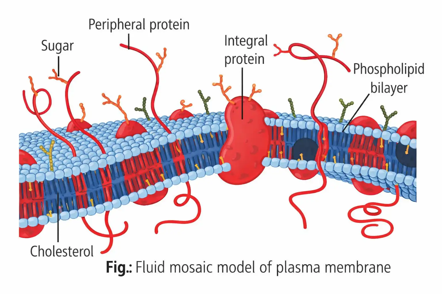
Plasma membrane — A selectively permeable membrane, mainly composed of phospholipids and proteins, that surrounds the cell.
Composition and structure
- Composition: Contains phospholipids, proteins, carbohydrates, water and cholesterol. Approximate proportions: lipids 20–79%, proteins 20–70%, carbohydrates 1–5% and water 20%.
- Bilayer: Phospholipid polar heads face the outer aqueous sides; hydrophobic hydrocarbon tails point inward.
- Proteins: Peripheral proteins lie on the membrane surface; integral proteins are partly or wholly embedded in it.
- Fluid mosaic model: Singer and Nicolson proposed this model in 1972: proteins are like “icebergs in a sea of lipids” within a phospholipid bilayer.
- Fluidity: The quasi-fluid bilayer permits lateral movement of proteins. This supports cell growth, junction formation, secretion, endocytosis and cell division.
| Transport | Direction / energy | Example |
|---|---|---|
| Simple diffusion | Neutral solutes move down the concentration gradient, from higher to lower concentration; no energy is required. | Neutral solute movement. |
| Osmosis | Diffusion of water across a selectively permeable membrane. | Water movement. |
| Carrier-mediated transport | Polar molecules use membrane carrier proteins because they cannot cross the non-polar lipid bilayer directly. | Transport of polar molecules. |
| Active transport | Substances move against the concentration gradient, from lower to higher concentration, using ATP. | pump. |
| Model | Main proposal |
|---|---|
| Danielli–Davson model (1935) | First lamellar model: two phospholipid layers and two protein layers. |
| Robertson model (1959) | A lipid bilayer sandwiched between protein layers on both sides. |
| Singer–Nicolson model (1972) | Fluid mosaic model: proteins are embedded in or associated with a fluid phospholipid bilayer. |
Cell Wall
Cell wall — A non-living, rigid, protective and semi-transparent outer covering of plant cells, fungi and some protists.
Composition, layers and functions
- Composition: Plant walls contain cellulose, hemicellulose, pectins and proteins. Algal walls may contain cellulose, galactans, mannans and minerals such as calcium carbonate.
- Primary wall: First-formed wall of a young plant cell; it can grow, but this capacity gradually diminishes as the cell matures.
- Secondary wall: Forms on the inner side of the wall in some mature cells that have stopped growing, e.g. tracheids, vessel elements and fibres.
- Middle lamella: Mainly calcium pectate; it holds neighbouring cells together. Fruit softens during ripening as this layer dissolves.
- Plasmodesmata: Protoplasmic connections through the cell wall and middle lamella link neighbouring cells; discovered and named by Strasburger.
- Structure and functions: Microfibrils are structural elements. The wall gives shape and rigidity, protects protoplasm from mechanical injury and undesirable macromolecules, and counteracts osmotic pressure to prevent bursting from excessive endosmosis.
Endomembrane System
Endomembrane system — A coordinated group of membrane-bound organelles that processes, sorts and recycles proteins and directs membrane traffic.
- Components: Endoplasmic reticulum, Golgi complex, lysosomes and vacuoles.
- Nuclear envelope: Also included because its lumen is continuous with the ER lumen.
Endoplasmic Reticulum
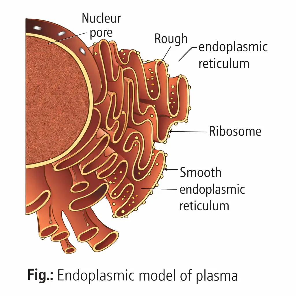
Endoplasmic reticulum (ER) — A cytoplasmic network of membrane-bound tubules and sacs that divides intracellular space into luminal and cytoplasmic compartments.
| Structure / type | Features |
|---|---|
| Cisternae | Flattened structures found in cells with synthetic roles. |
| Tubules | Involved in lipid and sterol synthesis. |
| Vesicles | Membrane-bound vacuolar structures. |
| Rough ER (RER) | Cisternae with ribosomes attached to the cytoplasmic surface. |
| Smooth ER (SER) | Mainly tubules and vesicles; lacks attached ribosomes. |
Functions
- RER: Actively synthesises and secretes proteins; bears enzymes for glycosylation and enzyme precursors used by the Golgi in lysosome formation.
- SER: Major site of lipid synthesis; in animal cells it synthesises steroid hormones and helps detoxify drugs and xenobiotics.
Golgi Apparatus
Structure and distribution
- Observation: Camillo Golgi first observed Golgi bodies in 1898 in nerve cells of barn owl and cat.
- Occurrence: Present in eukaryotic cells, but absent from male gametes of bryophytes and pteridophytes, mammalian red blood cells, plant sieve tubes and fungal cells.
- Arrangement: Animal cells usually have one Golgi apparatus or one connected complex. In plant cells, separate units are called dictyosomes.
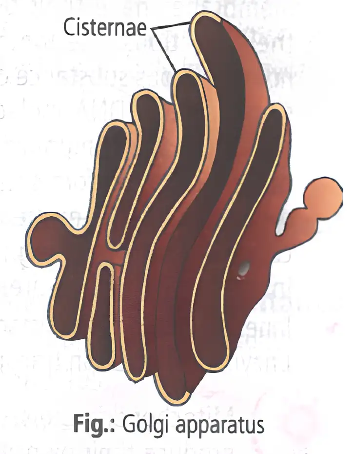
- Parts: Cisternae, vesicles, tubules and vacuoles.
- Cisternae: Flat, disc-shaped sacs about – in diameter, stacked parallel to each other. The stack has a convex cis (forming) face and a concave trans (maturing) face; the faces differ but are interconnected.
- Direction of traffic: ER-derived vesicles fuse with the cis face; materials move towards the trans face.
Functions
- Packaging: Materials are packaged and stored in vesicles released from the trans face.
- Protein processing: Proteins made on ER-bound ribosomes are modified in Golgi cisternae before release.
- Acrosome: Golgi vesicles help form the acrosome during spermatogenesis.
- Glycosylation: Addition of carbohydrate groups to proteins and lipids forms glycoproteins and glycolipids involved in cell recognition and adhesion.
Lysosomes
Lysosome — A small, single-membrane vesicle containing digestive hydrolytic enzymes, formed by packaging in the Golgi apparatus.
Properties and forms
- Discovery: First reported by Christian de Duve in 1955.
- Enzymes: Acid hydrolases—including lipases, proteases and carbohydrases—work best at acidic pH.
- Suicidal bags: Lysosomes are so called because their digestive enzymes can break down cellular material.
- Distribution: Present in animal cells except red blood cells; in plants and fungi, vacuoles take over lysosomal functions.
- Polymorphism: Different forms in a cell are primary lysosomes, secondary lysosomes, residual bodies and autophagic vacuoles.
Vacuoles
Vacuole — A membrane-bound space in the cytoplasm containing water, cell sap, excretory products and other materials.
Types and roles
- Tonoplast: The single membrane surrounding a vacuole; it transports ions and other substances into the vacuole, including against concentration gradients.
- Plant cells: Vacuoles can occupy up to 90% of cell volume and help maintain osmotic pressure and turgidity.
- Types: Sap, food, contractile and air vacuoles.
- Examples: Amoeba uses its contractile vacuole for osmoregulation and excretion; protists form food vacuoles by engulfing food particles.
Mitochondria
Structure and function
- Names and occurrence: Kolliker first observed mitochondria in 1857; Benda gave them this name in 1898. Present in aerobic eukaryotes; absent from prokaryotes and mature red blood cells.
- Role: Called the “powerhouse of the cell” because they carry out cellular respiration and generate energy.
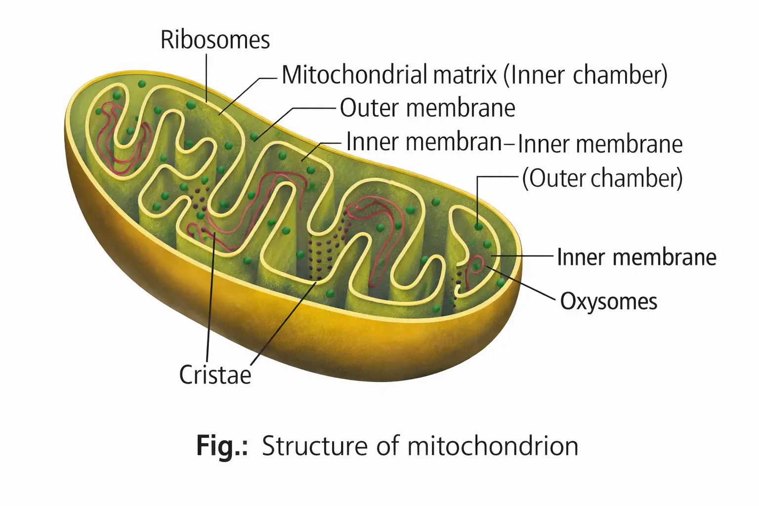
- Shape and size: Cylindrical or sausage-shaped; about – long and – wide (average diameter about ). They divide by fission.
- Compartments: The outer and inner membranes divide the organelle into outer and inner aqueous compartments. The inner compartment contains the matrix.
- Cristae: The inner membrane folds inward to form cristae, increasing surface area; each membrane has enzymes associated with its functions.
- Matrix: Contains a circular DNA molecule, some RNA, ribosomes and components for protein synthesis.
- Energy machinery: Inner membrane and cristae bear mushroom-like – particles (oxysomes); electron-transport-chain enzymes occur in the inner membrane in association with them.
Semi-autonomous nature
- Why semi-autonomous: Mitochondria have their own DNA, can replicate it, produce mRNA, tRNA and rRNA, and synthesise some of their proteins.
- Origin of new mitochondria: They arise from pre-existing mitochondria by fission.
Plastids
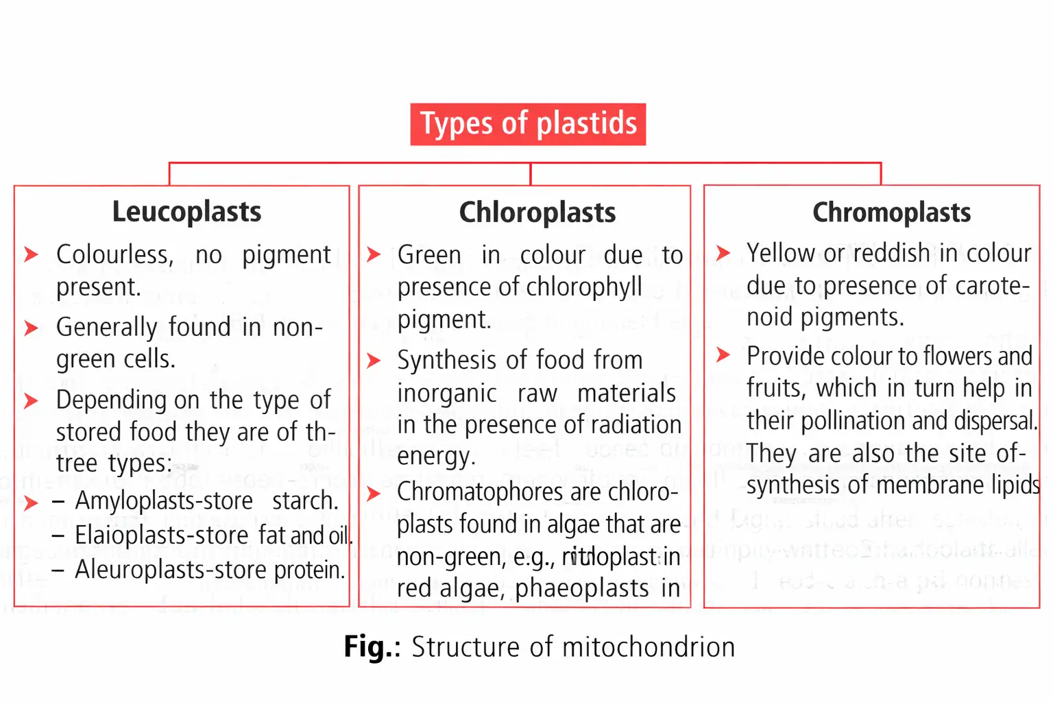
Plastids — Pigment-bearing organelles found in all plant cells and in euglenoids; their pigments impart specific colours to plants.
| Type | Pigment / appearance | Main feature |
|---|---|---|
| Chloroplast | Green; contains chlorophyll. | Photosynthetic plastid. |
| Chromoplast | Contains non-green coloured pigments. | Gives colour to plant parts. |
| Leucoplast | Colourless. | Storage plastid. |
Chloroplast structure
- Occurrence and size: Mainly in leaf mesophyll cells; shapes include lens-like, oval, spherical, discoid and ribbon-like. They are about – long and – wide. There is one per Chlamydomonas cell and about 20–40 per leaf mesophyll cell.
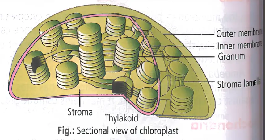
- Envelope: Double-membrane-bound; the inner membrane is relatively less permeable. The space enclosed by it is the stroma.
- Thylakoids and grana: Flattened membranous sacs called thylakoids are arranged in stacks called grana; intergranal thylakoids connect the stacks.
- Stroma lamellae: Flat membranous tubules connect thylakoids of different grana.
- Stroma contents: Enzymes for carbohydrate and protein synthesis, small double-stranded circular DNA molecules and ribosomes.
- Pigments and ribosomes: Chlorophyll is present in thylakoids. Chloroplast ribosomes are smaller () than cytoplasmic ribosomes ().
- Semi-autonomous nature: Like mitochondria, chloroplasts have DNA, ribosomes, RNA and organic molecules and can synthesise some of their proteins.
| Feature | Both organelles |
|---|---|
| Organisation | Semi-autonomous and enclosed by two membranes; the outer is smooth and permeable, while the inner is infolded and semipermeable. |
| Genetic machinery | Contain circular DNA, three types of RNA and ribosomes. |
| Energy role | Energy-transducing organelles that form ATP by a similar mechanism. |
| Occurrence | Found in eukaryotes and absent from prokaryotes. |
Ribosomes
Ribosome — A non-membrane-bound particle made of RNA and proteins that is the site of polypeptide synthesis.
| Type | Subunits | Typical location |
|---|---|---|
| Prokaryotes; also mitochondria and chloroplasts. | ||
| Eukaryotic cytoplasm. |
- Discovery: George Palade first observed ribosomes as dense particles under an electron microscope in 1953.
- Sedimentation coefficient: is a measure related to particle size and density; ribosomal subunit values do not add arithmetically to the whole-ribosome value.
- Function: Ribosomes are the cell’s protein factories.
Cytoskeleton
Cytoskeleton — A network of protein filaments and tubules forming the internal structural framework of the cell.
- Types: Microfilaments, microtubules and intermediate filaments.
- Functions: Maintains cell shape; regulates organelle orientation and distribution, intracellular transport and cellular movement.
Cilia and Flagella
| Point | Cilia | Flagella |
|---|---|---|
| Size and movement | Short, oar-like structures that move the cell or surrounding fluid. | Longer structures mainly responsible for cell movement. |
| Structure | Membrane-covered outgrowth with an axoneme. | Membrane-covered outgrowth with an axoneme. |
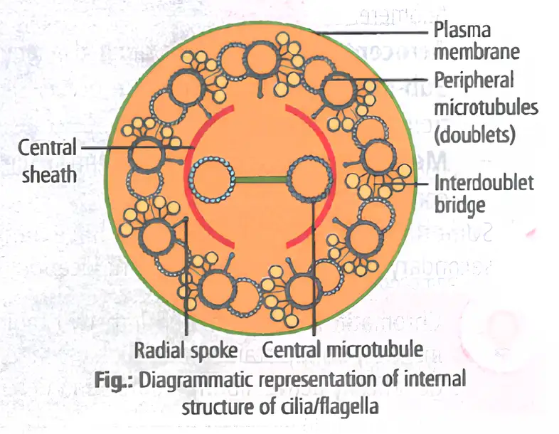
Axoneme
- Core arrangement: Usually nine peripheral microtubule doublets surround two central microtubules: the arrangement.
- Connections: A central sheath surrounds the central pair; nine radial spokes connect it to the peripheral doublets. Linkers connect neighbouring peripheral doublets.
- Base: Cilia and flagella emerge from centriole-like basal bodies.
- Proteins: Microtubules are made of tubulin; dynein arms and nexin links are associated with the axonemal structure.
- Prokaryotic flagella: Bacterial flagella are structurally different from eukaryotic flagella.
Centrosome and Centrioles
- Centrosome: Usually contains two cylindrical centrioles surrounded by amorphous pericentriolar material; the centrioles lie perpendicular to each other.
- Occurrence: Centrioles occur in nearly all animal cells and in motile plant cells, such as algal zoospores, fern sperm cells and motile algae.
- Structure: Each centriole has a cartwheel organisation with nine evenly spaced peripheral tubulin triplets. Adjacent triplets are linked; a protein hub connects to them by radial spokes.
- Functions: Centrioles form basal bodies of cilia and flagella and spindle fibres during animal-cell division.
Nucleus
Nucleus — The largest cell organelle, enclosed by a double membrane and containing the cell’s genetic information.
Nuclear structure and contents
- Discovery and name: Robert Brown described the nucleus in 1831; Flemming named the basic-dye-staining nuclear material chromatin.
- Interphase nucleus: Contains chromatin, nucleoplasm (nuclear matrix) and one or more nucleoli.
- Nuclear envelope: Two parallel membranes are separated by a perinuclear space of –. The outer membrane is continuous with the ER and may bear ribosomes.
- Nuclear pores: Form where the two envelope membranes fuse; they allow RNA and proteins to move in both directions between nucleus and cytoplasm.
- Nucleolus: A non-membranous region continuous with the nucleoplasm; it is the site of active rRNA synthesis. It is larger and more numerous in cells with high protein synthesis.
- Number of nuclei: Usually one per cell; Paramecium is binucleate, while some fungi and algae are multinucleate. Mature mammalian red blood cells and plant sieve-tube elements lack a nucleus.
Chromatin and chromosomes
- Chromatin: Loose, indistinct nucleoprotein fibres in interphase; contains DNA, histone and non-histone proteins, and RNA. During cell division, it forms distinct chromosomes.
- DNA length: A human cell contains about of DNA distributed among 46 chromosomes.
- Centromere and chromatids: Each chromosome has a primary constriction, the centromere, which holds its two chromatids; disc-shaped kinetochores occur on either side.
- Secondary constriction: Some chromosomes have a constant, non-staining secondary constriction that produces a small satellite.
| Type | Centromere position |
|---|---|
| Telocentric | At the terminal end. |
| Acrocentric | Near one end, just inward from the telomere. |
| Sub-metacentric | Slightly away from the middle. |
| Metacentric | In the middle. |
| Feature | Euchromatin | Heterochromatin |
|---|---|---|
| Packing and staining | Loosely packed; lightly stained. | Highly condensed; darkly stained. |
| Genetic activity | Genetically active; transcription occurs here. | Genetically inactive. |
Microbodies
Microbodies — Minute, membrane-bound vesicles containing various enzymes, found in both plant and animal cells.
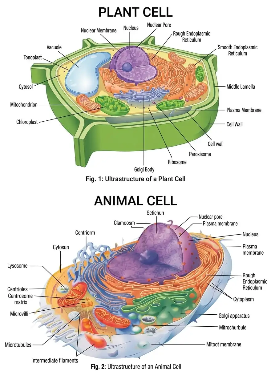
Plant Cell vs Animal Cell
| Feature | Plant cell | Animal cell |
|---|---|---|
| Cell wall | Present. | Absent. |
| Plastids | Present. | Absent. |
| Vacuole | Usually a large central vacuole. | No large central vacuole. |
| Centrioles | Generally absent; occur in motile plant cells. | Present. |
What are the main mechanisms of transport across the plasma membrane?
State the key structural features shared by mitochondria and chloroplasts.
Describe the axonemal arrangement of a eukaryotic cilium or flagellum.
Quick recap
- Eukaryotic cells have a membrane-bound nucleus, chromosomes and membrane-bound organelles.
- The fluid mosaic membrane is selectively permeable; diffusion and osmosis are passive, while active transport uses ATP.
- The endomembrane system coordinates the ER, Golgi, lysosomes and vacuoles in processing and transport.
- Mitochondria and chloroplasts are semi-autonomous organelles with circular DNA and ribosomes.
- Cilia and flagella usually have a axoneme; centrioles have nine peripheral triplets.
- Chromosomes are classified by centromere position; euchromatin is active and heterochromatin is inactive.
- Plant cells characteristically have a cell wall, plastids and a large central vacuole; animal cells have centrioles.
NEET Botany — ALL CHAPTERS
1
The Living World
2
Plant Kingdom
3
Morphology of Flowering Plants
4
Anatomy of Flowering Plants
5
The Unit of Life
6
Photosynthesis in Higher Plants
7
Respiration in Plants
8
Plant Growth and Development
9
Sexual Reproduction in Flowering Plants
10
Principles of Inheritance and Variation
11
Molecular Basis of Inheritance
12
Biotechnology :Principles and Processes
13
Biotechnology and its Applications