NEET UG . NEET Botany . Botany . Photosynthesis in Higher Plants
Chapter 6: Photosynthesis in Higher Plants
Ch 6
NEET Botany
NEET UG
What Do We Know
Photosynthesis and Its Importance
Food Relationships and Importance
- Autotrophs: Green plants make the food they need through photosynthesis.
- Heterotrophs: Other organisms depend directly or indirectly on green plants for food.
- Importance: Photosynthesis is the primary source of food on Earth and releases oxygen into the atmosphere.
- Energy source: Plants use sunlight; this use of solar energy supports life on Earth.
Photosynthesis — An enzyme-regulated anabolic process that manufactures organic compounds from carbon dioxide and water in chlorophyll-containing cells, using sunlight as the energy source.
Requirements and Starch Evidence
- Requirements shown: Chlorophyll, light and carbon dioxide are necessary for photosynthesis; water is also a reactant.
- Starch finding: Starch formed only in the green parts of leaves that were exposed to light.
- Conclusion: Photosynthesis occurs in chlorophyll-containing parts when light is available.
Quick recap
- Green plants are autotrophs; organisms that depend on them for food are heterotrophs.
- Photosynthesis is an enzyme-regulated anabolic process that makes organic compounds from carbon dioxide and water.
- It uses sunlight and occurs in chlorophyll-containing cells.
- Chlorophyll, light and carbon dioxide are required for photosynthesis; water is a reactant.
- Starch forms only in the green parts of leaves exposed to light.
- Photosynthesis is the primary source of food on Earth and releases oxygen.
Early Experiment
Moll’s Half-Leaf Experiment
- CO₂ requirement: Moll’s half-leaf experiment showed that carbon dioxide is essential for photosynthesis.
Priestley’s Experiment
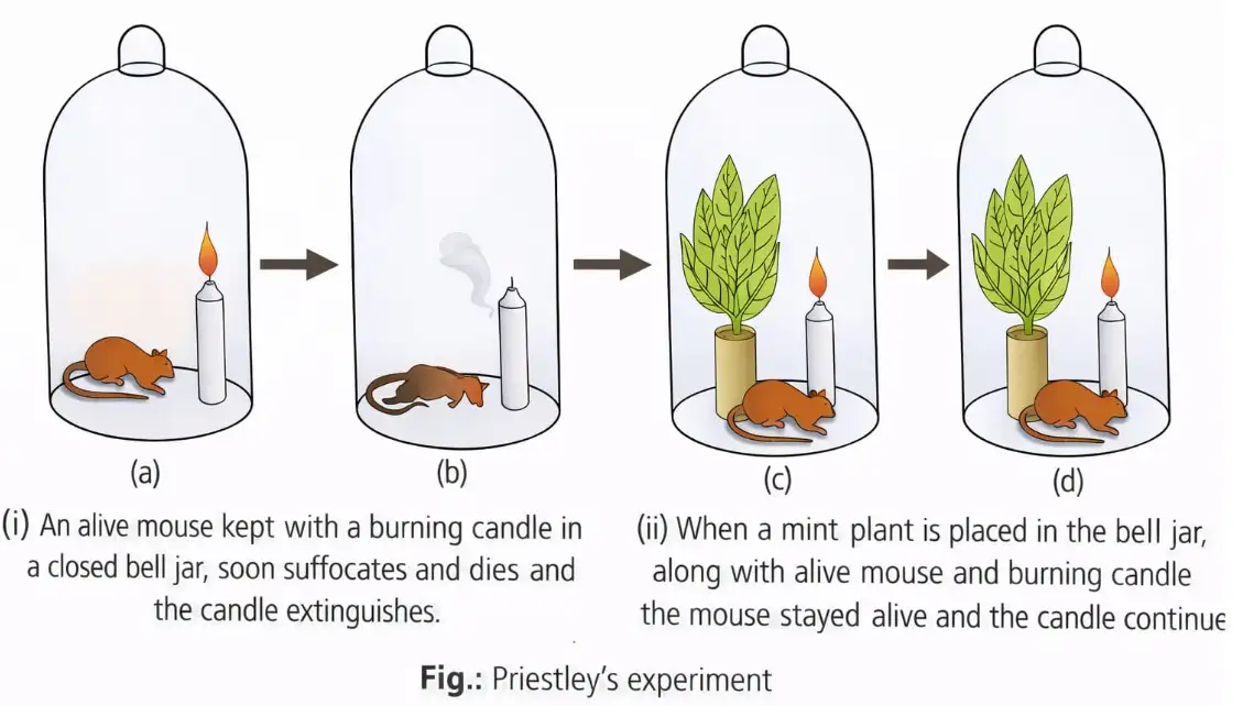
Milestones in Photosynthesis Research
- Moll: Removing prevents starch formation.
- Ingenhousz: Oxygen release by green plants occurs in sunlight.
- Van Niel and Ruben–Kamen: Oxygen released in photosynthesis originates from water.
Quick recap
- Moll’s half-leaf experiment showed that is necessary for starch formation in photosynthesis.
- Priestley proposed that plants restore air affected by animals and burning candles.
- Ingenhousz showed that green plants release oxygen in sunlight; von Sachs identified starch as the first visible product.
- Van Niel and Ruben–Kamen established that photosynthetic oxygen comes from water.
- Key later findings include the Calvin cycle, Emerson effect, Z-scheme, chemiosmotic theory and pathway.
- The source prints Engelmann’s date as 1884–1858 and Calvin’s as 1954–35; these dates are retained as printed.
Where Does Photosynthesis Take Place?
Photosynthesis and the Chloroplast
Chloroplast — A green plastid found in green parts of plants and the actual site of photosynthesis.
Location and Structure
- Main location: Photosynthesis occurs mainly in green leaves, especially in mesophyll cells, which contain many chloroplasts; it also occurs in other green plant parts.
- Position for light: Chloroplasts usually align along the walls of mesophyll cells to receive an optimum quantity of incident light.
- Shape and size: Higher-plant chloroplasts are usually discoid, ellipsoidal or biconvex lens-shaped; they are – long and – broad.
- Envelope: A double membrane surrounds each chloroplast and selectively regulates metabolite movement into and out of it.
- Stroma: The envelope encloses a hyaline, slightly electron-dense, granular, proteinaceous matrix containing the enzymes needed for photosynthesis.
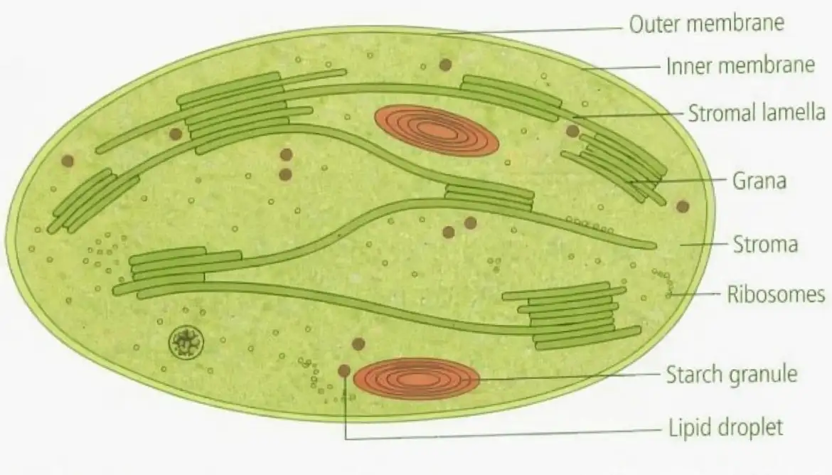
Chloroplast Regions and Photosynthetic Reactions
| Point | Light reactions | Dark reactions |
|---|---|---|
| Location | Thylakoid membrane system | Stroma |
| Energy relationship | Directly driven by light | Not directly driven by light; depend on ATP and NADPH from light reactions |
| Main role | Trap light energy and synthesise ATP and NADPH | Incorporate into the plant and lead to sugar synthesis; sugar can form starch |
“Dark reactions” does not mean they occur only in darkness; the name distinguishes reactions not directly driven by light.
Thylakoids, Grana and Photosystems
Thylakoid Membrane System
- Thylakoids: Flattened, sac-like lamellae within the stroma.
- Grana: Stacks formed by thylakoids.
- Stroma lamellae: Membranous connections between grana; also called fret channels.
- Photosystems: Light-reaction pigments and other factors cluster in thylakoid membranes to form photosystems.
| Photosystem | Location in chloroplast |
|---|---|
| Photosystem I (PSI) | Exclusively in stroma thylakoids and the non-appressed, stroma-facing regions of granal thylakoids |
| Photosystem II (PSII) | Appressed or partition regions of granal thylakoids |
Where are PSI and PSII located?
Quick recap
- Chloroplasts in green plant parts are the sites of photosynthesis; leaf mesophyll cells contain many of them.
- Chloroplasts align along mesophyll cell walls to receive an optimum quantity of incident light.
- A double-membrane envelope surrounds the enzyme-containing stroma.
- Light reactions occur in thylakoid membranes and produce ATP and NADPH.
- Stromal reactions use ATP and NADPH to incorporate and form sugars, which may form starch.
- Thylakoids stack into grana, interconnected by stroma lamellae (fret channels).
- PSI is associated with stroma thylakoids and non-appressed granal regions; PSII with appressed granal regions.
How Many Types of Pigments are Involved in Photosynthesis?
Photosynthetic pigments — Pigments absorb light for photosynthesis; leaf colour comes from several pigments, not a single pigment.
| Pigment | Typical colour |
|---|---|
| Chlorophyll | Bright or blue-green |
| Chlorophyll | Yellow-green |
| Xanthophylls | Yellow |
| Carotenoids | Yellow to yellow-orange |
Chlorophylls
Types and roles
- Types: Plants other than bacteria have chlorophylls , , , and ; higher plants have only chlorophylls and in their chloroplasts.
- Chlorophyll : Makes up roughly one-fourth of the total chlorophyll content.
- Chlorophyll : The universal photosynthetic pigment in photosynthetic plants except bacteria; it carries out the primary reaction that converts light energy into chemical or electrical energy.
- Accessory pigments: Chlorophyll and other pigments absorb light at additional wavelengths and transfer the absorbed energy to chlorophyll through electron spin resonance.
- Reaction centres: Some chlorophyll molecules, including and , separate electrical charges and convert light energy into electrical energy.
Chlorophyll structure
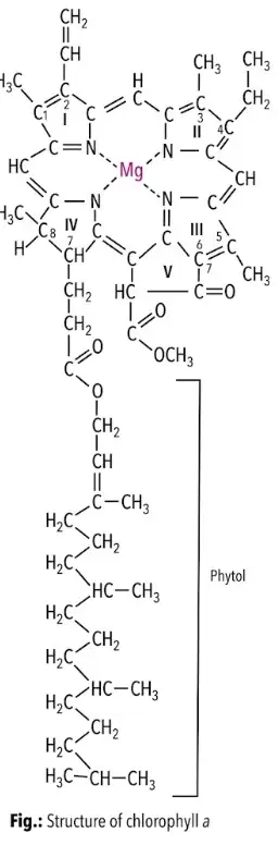
- Configuration: Chlorophyll has a tadpole-like structure with a porphyrin head and a phytol tail.
- Porphyrin head: Contains four pyrrole rings linked by methine bridges (); each ring has four carbon atoms and one nitrogen atom.
- Magnesium: A non-ionic magnesium atom sits at the centre, held by pyrrole nitrogens through two covalent and two coordinate bonds.
- Phytol tail: This insoluble long-chain alcohol () anchors chlorophyll in the lipid part of the thylakoid membrane.
- Structure studied: Willstätter, Stoll and Fischer studied chlorophyll structure in 1912.
Carotenoids
Types and properties
- Occurrence: Carotenoids are yellow, brown or reddish pigments associated with chlorophylls in chloroplasts; they also occur in chromoplasts.
- Role: Along with chlorophyll , carotenoids act as accessory pigments by transferring absorbed energy to chlorophyll .
- Carotenes: Orange hydrocarbons with general formula . The commonest is -carotene, which animals and humans convert to vitamin A.
- Lycopene: This carotene gives tomatoes and chillies their red colour.
- Xanthophylls: Yellowish, oxygen-containing carotene derivatives; examples are cryptoxanthin (), lutein and zeaxanthin ().
- Solubility: Both groups dissolve in organic solvents such as chloroform and ethyl ether; carotenes are more soluble in carbon disulphide than xanthophylls.
| Type | Key feature | Examples |
|---|---|---|
| Carotenes | Orange hydrocarbons; | -carotene, lycopene |
| Xanthophylls (carotenols) | Yellow, oxygen-containing derivatives | Cryptoxanthin, lutein, zeaxanthin |
Phycobilins
- Occurrence: Important accessory pigments in blue-green algae (cyanobacteria), cryptomonads and red algae.
- Form and location: Protein-linked, water-soluble pigments in small granules called phycobilisomes, attached to thylakoids.
- Structure: Open tetrapyrrole pigments that lack both magnesium and a phytol chain.
- Types: Phycocyanin and phycoerythrin.
Action and Absorption Spectra
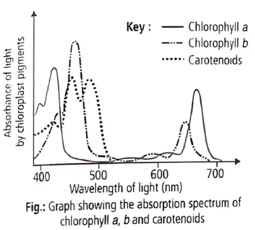
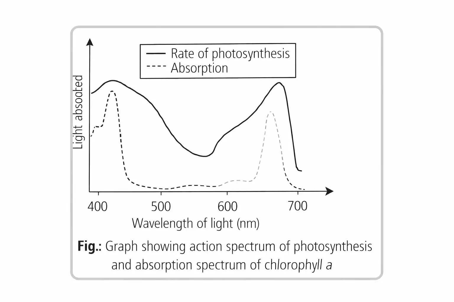
Absorption spectrum — A graph showing the amount of energy absorbed by a substance at different wavelengths of light.
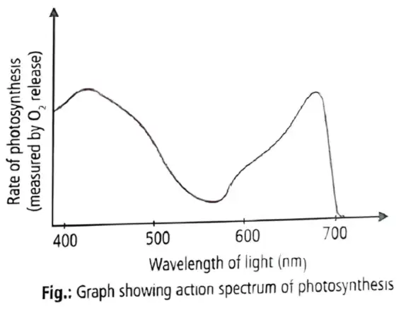
Action spectrum — A graph showing the relative rate of photosynthesis at different wavelengths of light.
What the spectra show
- Absorption peaks: Chlorophylls and absorb most strongly in the blue and red regions, where photosynthesis is also fastest.
- Correspondence: The action spectrum closely matches the absorption spectra of chlorophylls and .
- Green light: Photosynthesis is lowest in green light, but it also occurs to some extent at other visible wavelengths.
- Wider light capture: Accessory pigments broaden the range of wavelengths used in photosynthesis by transferring absorbed energy to chlorophyll .
- Protection: Accessory pigments help protect chlorophyll from photo-oxidation.
What is the difference between an absorption spectrum and an action spectrum?
Why are chlorophyll and carotenoids called accessory pigments?
Quick recap
- Leaf colour comes from chlorophyll , chlorophyll , xanthophylls and carotenoids.
- Higher plants contain chlorophylls and ; chlorophyll is the primary pigment and chlorophyll is about one-fourth of total chlorophyll.
- Accessory pigments transfer absorbed energy to chlorophyll through electron spin resonance and protect it from photo-oxidation.
- Carotenoids include carotenes and xanthophylls; phycobilins occur in cyanobacteria, cryptomonads and red algae.
- Absorption spectra show light absorbed, while action spectra show photosynthesis rates at different wavelengths.
- Photosynthesis is strongest in blue and red light and weakest in green light.
What is Light Reaction?
Light Reactions and Photosystems
Light-Harvesting Complexes
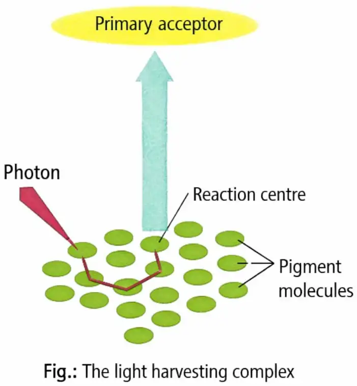
- Organisation: Pigments form two light-harvesting complexes, one in each of Photosystem I (PS I) and Photosystem II (PS II).
- Naming: PS I and PS II are named by order of discovery, not by the order in which they function.
- Antenna: In each photosystem, all pigments except one chlorophyll a molecule form the light-harvesting antenna.
- Efficiency: Different antenna pigments absorb different wavelengths, increasing the light captured for photosynthesis.
Reaction Centres
- Reaction centre: The single chlorophyll a molecule in each photosystem acts as its reaction centre.
- PS I: Its reaction-centre chlorophyll absorbs maximally at and is called .
- PS II: Its reaction-centre chlorophyll absorbs maximally at and is called .
| Feature | PS I | PS II |
|---|---|---|
| Pigments | More chlorophyll a; comparatively less chlorophyll b and carotenoids | Equal amounts of chlorophyll a and chlorophyll b; more carotenoids than PS I |
| Reaction centre and core | and the core antenna are bound to proteins Psa A and Psa B | The reaction-centre core contains membrane proteins D₁ and D₂ |
| Other components | Three membrane-associated iron–sulphur proteins (Fe-S centres), also known as bound ferredoxins | , additional chlorophylls, carotenoids, pheophytins and plastoquinones are bound to D₁ and D₂; includes cytochrome b₆f complex and plastocyanin |
| Photophosphorylation | Participates in both cyclic and non-cyclic photophosphorylation | Participates only in non-cyclic photophosphorylation, in association with PS I |
Emerson’s Enhancement Effect
Red Drop and Enhancement
- Emerson (1957): Photosynthesis sharply declines when monochromatic light with wavelength above is used alone; this is the red drop.
- Shorter wavelengths: Supplied alone, these wavelengths were also ineffective in photosynthesis.
- Enhancement effect: Photosynthesis increases when shorter- and longer-wavelength light are supplied simultaneously.
- Cause: Different harvesting molecules surround a trap centre, and two interconnected pigment systems contain some common pigments.
- PS I is ; PS II is .
- PS I supports cyclic and non-cyclic photophosphorylation; PS II supports only non-cyclic photophosphorylation.
- Using shorter and longer wavelengths together increases photosynthesis compared with either wavelength range alone.
Quick recap
- Light reactions absorb light, split water, release oxygen, and form ATP and NADPH.
- Each photosystem has an antenna of pigments and one chlorophyll a reaction-centre molecule.
- PS I has the centre; PS II has the centre.
- PS I participates in cyclic and non-cyclic photophosphorylation; PS II only in non-cyclic photophosphorylation.
- Emerson’s red drop is the sharp decrease in photosynthesis under monochromatic light above .
- Simultaneous shorter- and longer-wavelength light increases photosynthesis because the two pigment systems cooperate.
The Electron Transport
Non-cyclic Photophosphorylation and the Z Scheme
Phosphorylation — The cellular synthesis of ATP, which stores energy in its chemical bonds.
Non-cyclic electron flow
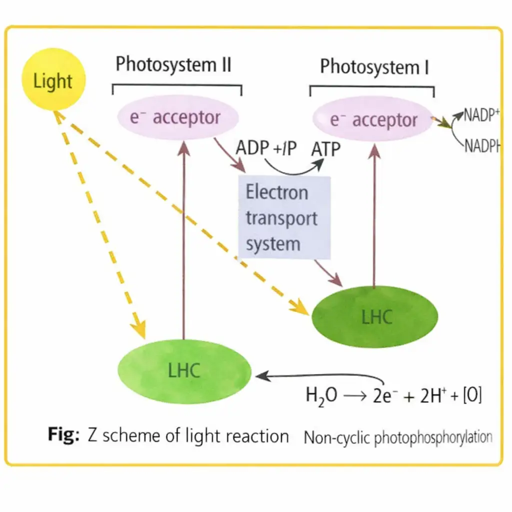
- Photosystem order: acts first, followed by ; the photosystems are connected by an electron transport chain.
- Products: Electron flow through both photosystems produces ATP and reduces to .
- At : Light at excites reaction-centre chlorophyll ; its electrons move to an acceptor and then downhill through cytochromes.
- At : Electrons arrive from and replace those lost by . Light at excites electrons, which ultimately reduce .
- Z scheme: The electron pathway rises with light-driven excitation and falls as electrons pass downhill through carriers; its redox-potential plot resembles the letter Z.
Non-cyclic electron pathway
- Light excites in → electrons reach a primary acceptor.
- Electrons pass downhill through the electron transport chain → they reach .
- Light excites → its electrons pass to another acceptor and finally reduce to .
Splitting of Water
- Photolysis: Water splits at the water-splitting complex associated with , replacing the electrons removed from .
- Location: The complex lies on the inner, lumen-facing side of the thylakoid membrane; released protons accumulate in the lumen.
- Products: Water splitting supplies electrons and protons and releases oxygen, a net product of photosynthesis.
- Electron replacement: Electrons from are passed on to replace those lost by .
Cyclic Photophosphorylation
Cyclic photophosphorylation — A light-driven process in which electrons from excited return to the same reaction centre after passing through electron carriers.
| Feature | Cyclic | Non-cyclic |
|---|---|---|
| Photosystems | only | followed by |
| Electron path | Electrons return to | Electrons ultimately reduce |
| Main products | ATP; no or is produced by this flow | ATP, and oxygen from water splitting |
| Favoured conditions | Low light intensity, wavelength longer than , or inhibited fixation | Both photosystems operate in sequence |
Cyclic electron pathway
- Excitation: A photon excites the photocentre; the expelled electron gains about of energy, leaving the photocentre oxidised.
- Carrier sequence: The electron passes through plastoquinone (), the complex, ferredoxin (), cytochrome and plastocyanin, then returns to .
- Outcome: The cyclic flow produces ATP but does not produce or oxygen.
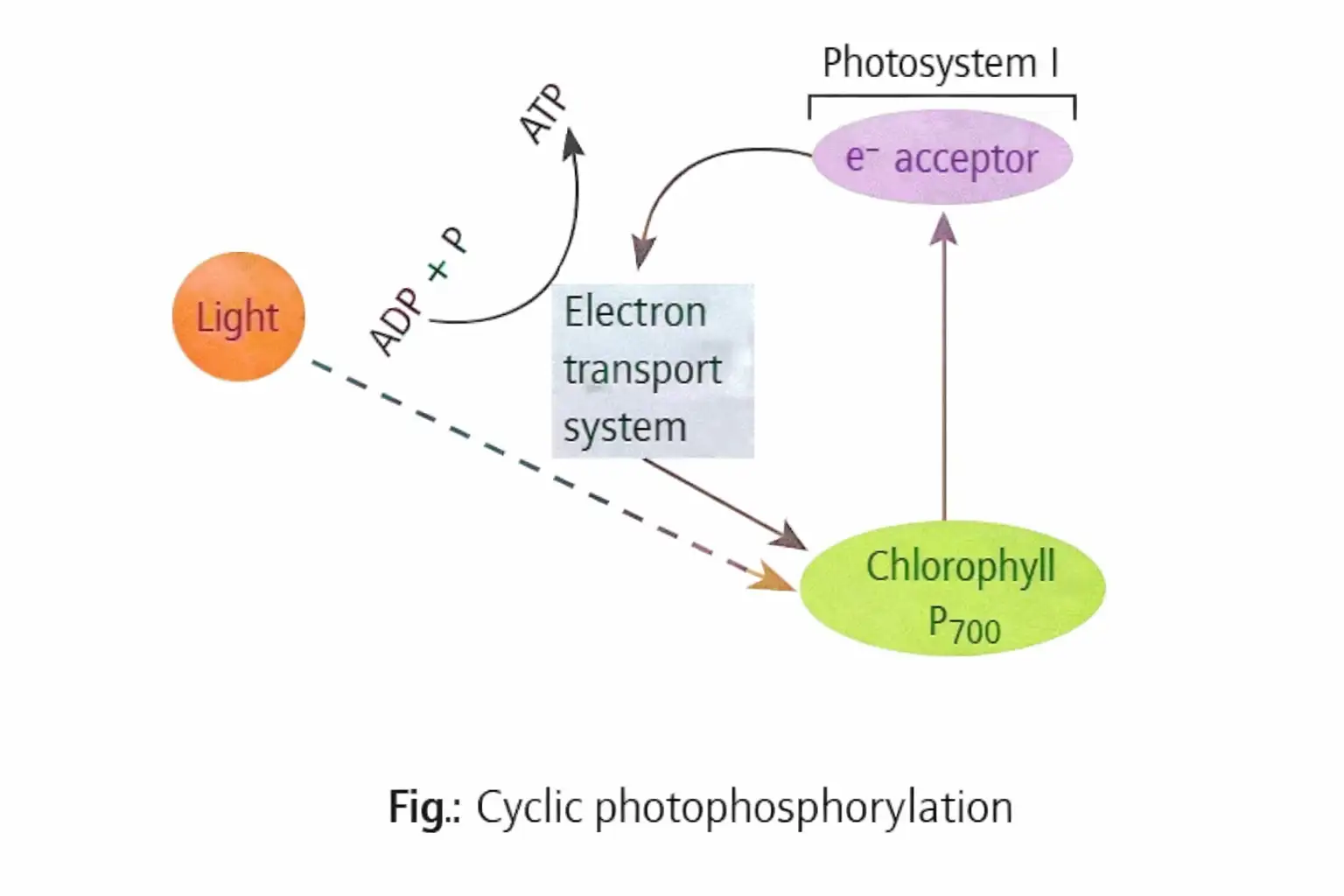
Chemiosmotic Hypothesis
Chemiosmotic hypothesis — Peter Mitchell proposed in 1961 that ATP synthesis is driven by a proton gradient across the thylakoid membrane.
How the proton gradient forms and drives ATP synthesis
- Water splits on the lumen side of the membrane → released accumulates in the thylakoid lumen.
- Electron transport moves protons from the stroma into the lumen through an H-carrier mechanism.
- Reduction of on the stroma side uses stromal protons → proton concentration falls in the stroma and rises in the lumen.
- The resulting gradient creates a lower pH in the lumen → protons flow back to the stroma through the channel.
- Proton flow provides energy for a conformational change in → ATP synthase forms ATP.
| Component | Location or role |
|---|---|
| Proton gradient | High concentration in the lumen relative to the stroma |
| Membrane-embedded portion of ATPase; forms the channel for facilitated proton diffusion to the stroma | |
| Projects from the thylakoid membrane into the stroma; its conformational change enables ATP synthesis | |
| Essential requirements | A membrane, proton pump, proton gradient and ATPase |
Importance
- Energy coupling: Proton movement down its gradient through ATPase releases energy that drives ATP synthesis.
- Use of products: ATP and produced in the light reactions are used in the stroma for fixation and sugar synthesis.
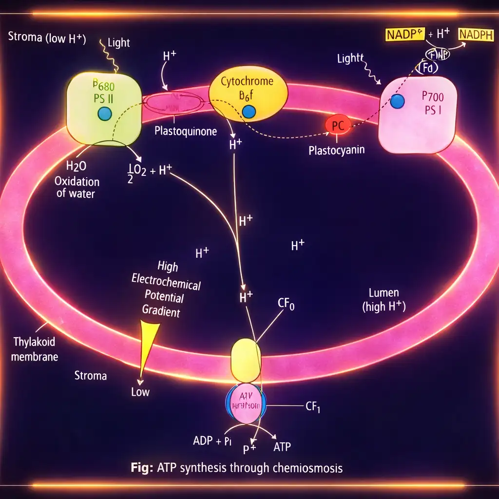
Do not confuse the pathways: cyclic flow uses only and returns electrons to ; non-cyclic flow uses both photosystems and transfers electrons to .
What is the Z scheme?
How does the proton gradient lead to ATP formation?
Quick recap
- Non-cyclic photophosphorylation uses then and produces ATP and .
- The Z scheme shows light-driven electron excitation and downhill electron transfer between carriers.
- Water splitting at the lumen-facing complex provides electrons, protons and oxygen.
- Cyclic photophosphorylation uses only; electrons return to and ATP is produced.
- Chemiosmosis relies on a proton gradient across the thylakoid membrane and proton flow through ATPase.
- forms the proton channel, while stromal-facing drives ATP synthesis using the gradient's energy.
Where are the ATP and NADPH Used?
Use of Light-Reaction Products
Biosynthetic or dark phase
- Light-reaction products: ATP, NADPH and O₂; O₂ diffuses out of the chloroplast, while ATP and NADPH drive sugar synthesis.
- Light dependence: This phase does not directly require light, but depends on ATP and NADPH made in the light reaction, as well as CO₂ and H₂O.
- Location: It occurs in the chloroplast stroma, where the required enzymes are present.
- Historical note: F.F. Blackman established the dark reaction in 1905; Calvin, Benson and Bassham later studied it in detail and received the Nobel Prize in 1961.
The Calvin Cycle
Calvin cycle — A series of enzyme-catalysed reactions that fixes CO₂ using ATP and NADPH and converts it into carbohydrate.
Core facts
- CO₂ acceptor: Ribulose-1,5-bisphosphate (RuBP), a five-carbon ketose sugar.
- First stable product: 3-phosphoglyceric acid (3-PGA), a three-carbon compound.
- Three phases: Carboxylation → reduction → regeneration of RuBP.
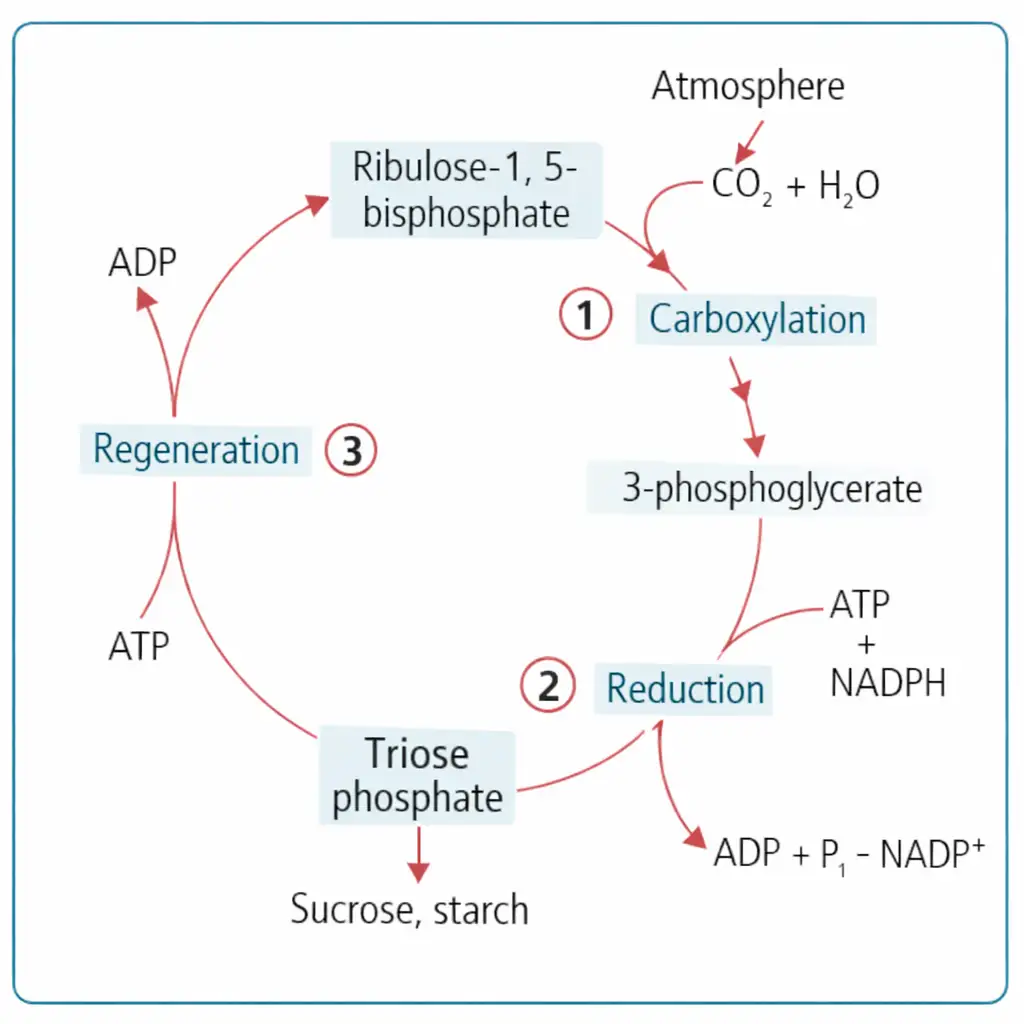
Carboxylation
- Enzyme: RuBP carboxylase-oxygenase (RuBisCO) catalyses the fixation of CO₂; it is the most abundant protein in the biological world.
- Reaction sequence: CO₂ combines with RuBP to form a short-lived intermediate, 2-carboxy-3-keto-1,5-bisphosphoribotol.
- Product formation: The intermediate immediately splits in the presence of water to form two molecules of 3-PGA.
Reduction
- Purpose: A series of reactions uses light-reaction products to reduce the fixed carbon and form carbohydrate.
- Per CO₂ fixed: The reduction steps use 2 ATP for phosphorylation and 2 NADPH for reduction.
- Glucose formation: Fixation of 6 CO₂, involving six turns of the cycle, is required to form one glucose molecule.
Regeneration of RuBP
- Role: RuBP must be regenerated so that the cycle can continue fixing CO₂.
- Energy cost: Regeneration uses 1 ATP per CO₂ fixed for phosphorylation.
The two ATP used in reduction plus one ATP in RuBP regeneration give the total requirement.
What are the three phases of the Calvin cycle, and what happens in each?
How much ATP and NADPH are required to fix one CO₂ molecule?
Quick recap
- ATP and NADPH from the light reaction power sugar synthesis in the chloroplast stroma.
- RuBP is the five-carbon CO₂ acceptor; 3-PGA is the first stable product.
- RuBisCO catalyses carboxylation, producing two 3-PGA molecules from the transient intermediate.
- The Calvin cycle proceeds through carboxylation, reduction and RuBP regeneration.
- Each CO₂ fixed requires 3 ATP and 2 NADPH; six CO₂ and six turns yield one glucose.
The C₄ Pathway
Pathway (Hatch–Slack Pathway)
pathway — A photosynthetic -fixation pathway in which the first stable product is a four-carbon compound; it is also called the Hatch–Slack pathway.
Plants and Kranz anatomy
- Examples: Maize, sugarcane, sorghum, *Atriplex* and *Amaranthus* are plants; they include both monocots and dicots.
- Kranz anatomy: Large bundle sheath cells surround the vascular bundles in a wreath-like arrangement, forming one or more layers.
- Mesophyll cells: Carry out light reactions, release and produce ATP and NADPH; they lack RuBisCO and contain PEP carboxylase.
- Mesophyll chloroplasts: Lack starch.
- Bundle sheath cells: Contain RuBisCO and have agranal chloroplasts; cyclic photophosphorylation can produce ATP.
- Bundle sheath reactions: Photolysis and evolution are absent; these cells are protected from oxygen released by mesophyll cells.
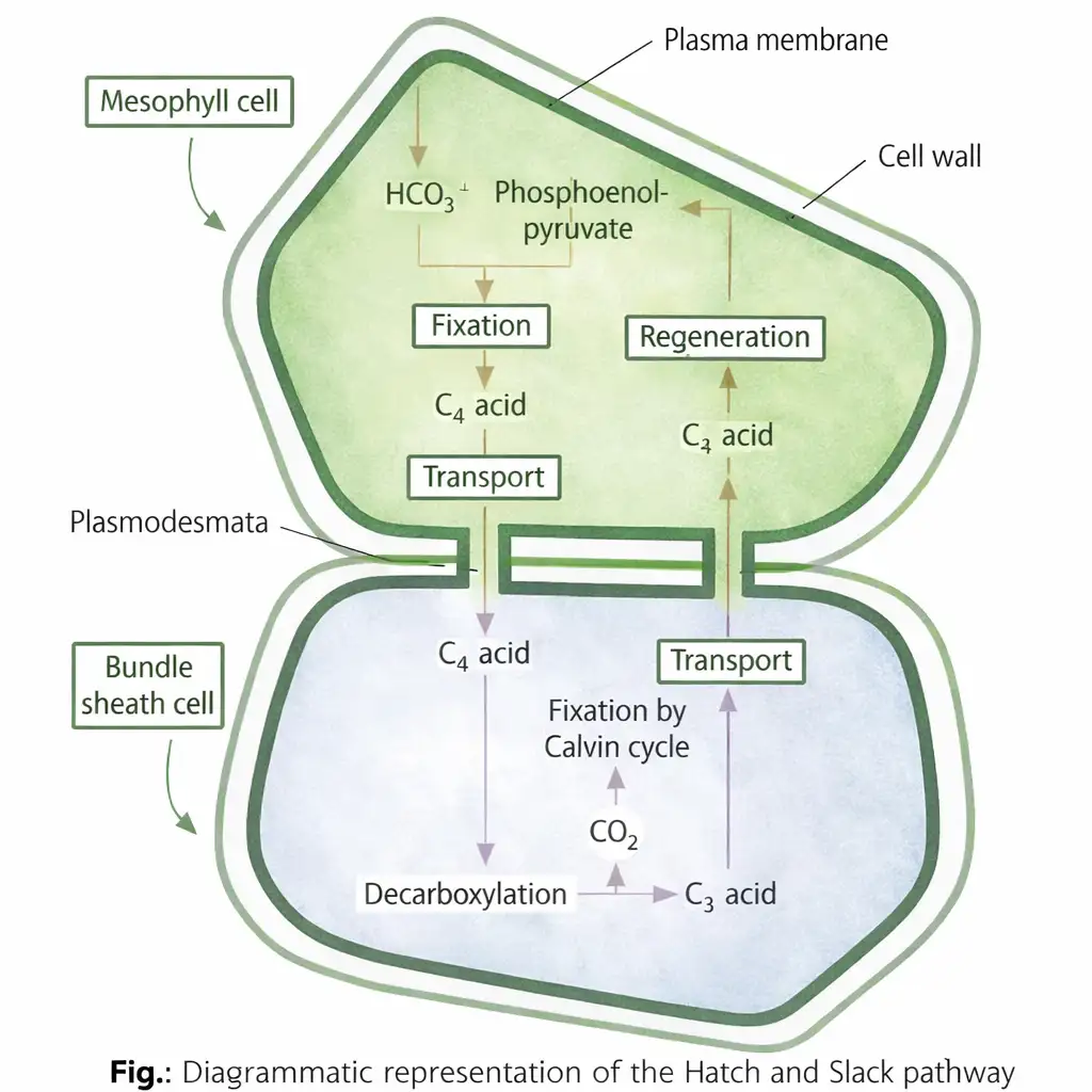
pathway: sequence of fixation
From initial fixation to the Calvin cycle
- In mesophyll cells, PEP is the initial acceptor; PEP carboxylase fixes to form oxaloacetate (OAA).
- OAA is reduced to malic acid or transaminated to form aspartic acid.
- Malic acid or aspartic acid moves to bundle sheath cells and is decarboxylated, producing pyruvate and .
- The released is fixed in the Calvin cycle by RuBisCO, with RuBP as the final acceptor.
- Initial fixation: PEP + PEP carboxylase in mesophyll cells → OAA, a four-carbon compound.
- Final fixation: RuBP + RuBisCO in bundle sheath cells → Calvin cycle.
and plants
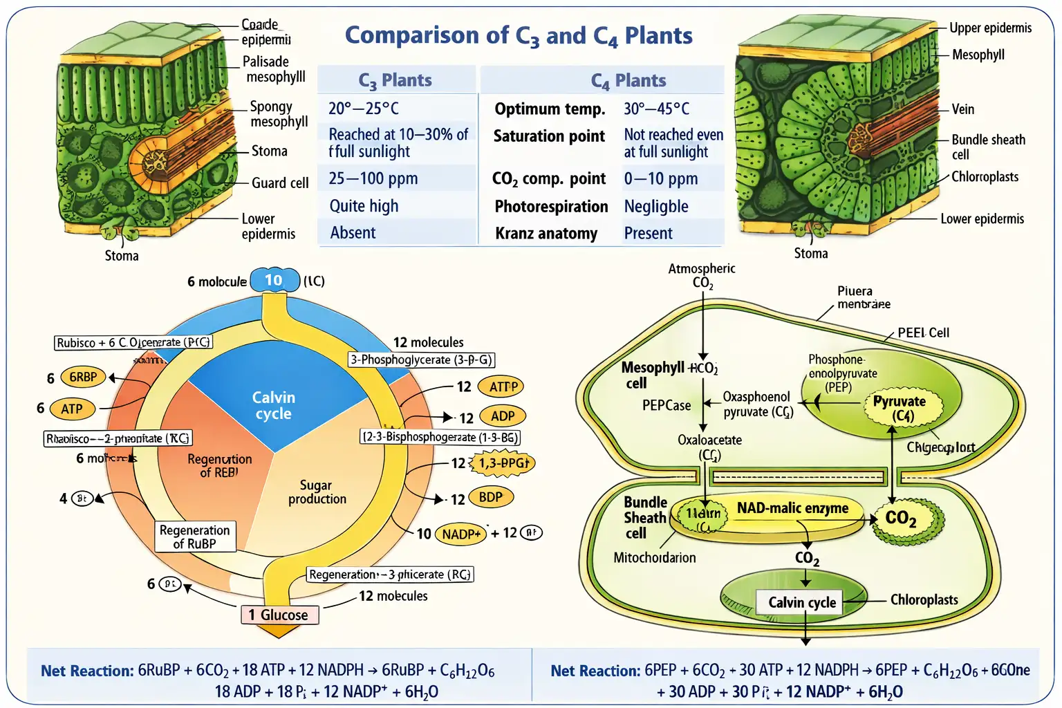
| Point | plants | plants |
|---|---|---|
| First stable product | Three-carbon compound | Four-carbon compound, initially OAA |
| Initial fixation | By RuBisCO in the Calvin cycle | By PEP carboxylase in mesophyll cells |
| Cell arrangement | Kranz anatomy absent | Kranz anatomy present; initial fixation and Calvin-cycle fixation occur in different cells |
CAM pathway
Crassulacean acid metabolism (CAM) — A photosynthetic mechanism with double fixation of , found in succulent plants such as those of the Crassulaceae family.
| Time | Stomata and fixation | Main events |
|---|---|---|
| Night | Stomata open; atmospheric enters. | PEP carboxylase fixes using PEP. Malic acid forms and is stored in cell vacuoles. |
| Day | Stomata closed; light is available. | Malic acid leaves the vacuoles and is decarboxylated by malic enzyme, producing pyruvate and releasing . RuBisCO fixes this in the Calvin cycle using RuBP. |
| Point | plants | CAM plants |
|---|---|---|
| Separation | Spatial: initial PEP carboxylase fixation and RuBisCO fixation occur in different cells. | Temporal: both fixation steps occur in the same cells at different times. |
| Timing of initial fixation | In mesophyll cells before transport to bundle sheath cells. | At night; the resulting malic acid is stored until daytime. |
| Water conservation | Bundle sheath cells concentrate released for the Calvin cycle. | Night-time opening and daytime closure of stomata reduce water loss. |
How do the pathway and CAM differ in separating the two stages of fixation?
Quick recap
- plants are named for their first stable product, a four-carbon compound; the pathway is also called Hatch–Slack.
- Kranz anatomy has large bundle sheath cells around vascular bundles; mesophyll cells use PEP carboxylase and bundle sheath cells contain RuBisCO.
- PEP fixes to form OAA, which becomes malic acid or aspartic acid and moves to bundle sheath cells.
- Decarboxylation in bundle sheath cells releases for the Calvin cycle; RuBP is its final acceptor.
- CAM plants fix at night into malic acid and use the released for the Calvin cycle during the day.
- plants separate fixation spatially, whereas CAM plants separate it temporally.
Photorespiration
Photorespiration: Site and Key Features
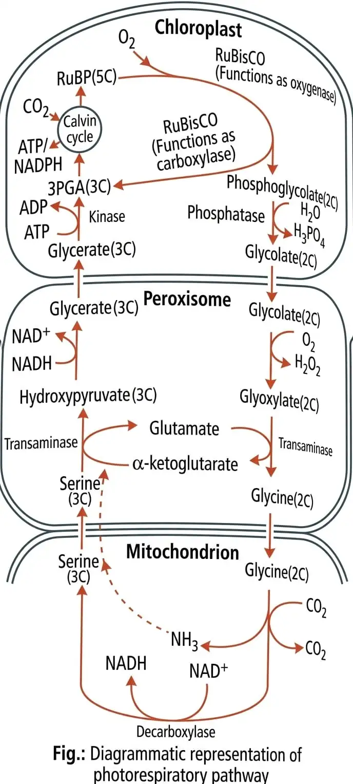
Photorespiration — A cyclic process in which RuBisCO uses oxygen to oxidise RuBP instead of fixing carbon dioxide.
- Site: The process occurs in the chloroplast; the peroxisome and mitochondrion are also required to complete it.
- Enzyme: RuBisCO (RuBP carboxylase-oxygenase) catalyses the process.
- CO₂ affinity: RuBisCO has high affinity for when the ratio is nearly equal.
- Conditions: At high temperature, RuBisCO functions as an oxygenase and oxidises RuBP instead of fixing carbon dioxide.
- Products: RuBP oxidation forms one 3-carbon phosphoglyceric acid molecule and one 2-carbon phosphoglycolate molecule.
- Energy and sugar: Photorespiration produces no ATP, NADPH or sugars.
Photorespiration in C₄ Plants
- CO₂ concentration: C₄ acids formed in mesophyll cells move to bundle sheath cells and release there.
- Effect on RuBisCO: The increased local concentration favours its carboxylase activity and minimises oxygenase activity.
- Photorespiration: It does not occur in C₄ plants because this mechanism prevents RuBisCO from acting as an oxygenase.
- Advantages: C₄ plants have better productivity and yields, and tolerate higher temperatures.
How C₄ Plants Prevent Photorespiration
- C₄ acid moves from mesophyll cells to bundle sheath cells.
- The C₄ acid breaks down and releases near RuBisCO.
- Higher local favours RuBisCO carboxylase activity over oxygenase activity.
- Photorespiration does not occur in C₄ plants.
Why does photorespiration not occur in C₄ plants?
What are the products and energy outcomes of photorespiration?
Quick recap
- Photorespiration is a cyclic process involving the chloroplast, peroxisome and mitochondrion.
- RuBisCO has high affinity for when the ratio is nearly equal; at high temperature it can act as an oxygenase.
- RuBP oxygenation produces 3-phosphoglyceric acid and phosphoglycolate.
- Photorespiration produces no ATP, NADPH or sugars.
- C₄ plants release near RuBisCO in bundle sheath cells, so photorespiration does not occur.
- C₄ plants consequently show better productivity and yield, and tolerate higher temperatures.
Factors Affecting Photosynthesis
Carbon Dioxide Concentration (CO₂)
CO₂ compensation point (threshold value) — The CO₂ concentration at which CO₂ fixed in photosynthesis equals CO₂ released by respiration and photorespiration, so illuminated plant parts have no net CO₂ exchange.
CO₂ effects
- Increasing CO₂: Raises photosynthesis in most C₃ plants up to about .
- Reduced CO₂: At the compensation point, illuminated plant parts stop taking up CO₂ from their surroundings.
| Plant type | Compensation point | CO₂ saturation |
|---|---|---|
| C₃ | – | Beyond |
| C₄ | – | About |
Light Intensity
Light compensation point — The low light intensity at which photosynthesis and respiration balance, producing no net gaseous exchange.
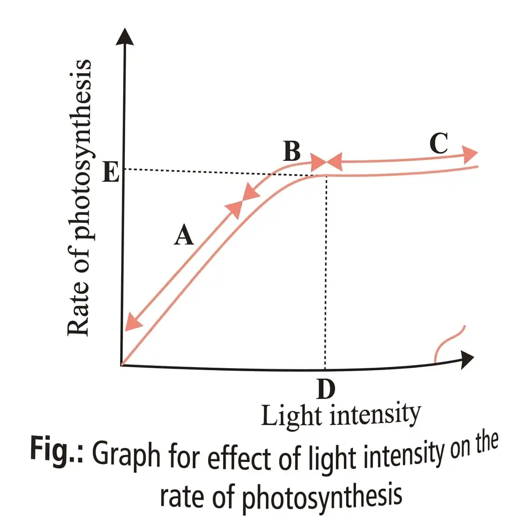
Light saturation point — The light intensity at which a plant reaches its maximum rate of photosynthesis.
Light effects
- Rising light: Photosynthesis increases with light intensity until the saturation point; saturation occurs at about of full sunlight.
- Excess light: Beyond a certain intensity, chlorophyll breaks down and photosynthesis decreases.
Temperature
Temperature effects
- Minimum to optimum: The photosynthesis rate doubles for every rise in temperature.
- Above optimum: The rate may rise briefly, then declines; this delayed decline is called the time factor.
- Climate adaptation: Tropical plants generally have a higher temperature optimum than temperate plants.
Oxygen
Warburg effect — The decline in photosynthesis rate caused by very high oxygen concentration; it occurs in all plants.
Water
- Amount used: Photosynthesis consumes only a very small amount of water.
- Water availability: The process is highly sensitive to dehydration.
Limiting Factor and Blackman’s Law
Limiting factor — A factor that is deficient enough that increasing its magnitude directly increases the rate of a process.
Law of limiting factors
- Blackman’s law: Proposed by F. F. Blackman in ; the rate of a process is limited by the factor that is least available or slowest to act.
- Basis: This law follows Liebig’s law of the minimum: when several factors control a process, its rate is limited by the slowest factor.
Quick recap
- CO₂ raises photosynthesis in most C₃ plants until saturation; compensation points differ between C₃ and C₄ plants.
- The CO₂ compensation point, also called the threshold value, is where fixation equals CO₂ release through respiration and photorespiration.
- Photosynthesis rises with light until saturation, while excessive light can damage chlorophyll and reduce the rate.
- Temperature raises the rate up to an optimum; above it, the rate eventually declines, showing the time factor.
- Very high oxygen inhibits photosynthesis (Warburg effect), and dehydration strongly affects the process.
- Under Blackman’s law, the least available factor limits the rate of photosynthesis.
NEET Botany — ALL CHAPTERS
1
The Living World
2
Plant Kingdom
3
Morphology of Flowering Plants
4
Anatomy of Flowering Plants
5
The Unit of Life
6
Photosynthesis in Higher Plants
7
Respiration in Plants
8
Plant Growth and Development
9
Sexual Reproduction in Flowering Plants
10
Principles of Inheritance and Variation
11
Molecular Basis of Inheritance
12
Biotechnology :Principles and Processes
13
Biotechnology and its Applications