NEET UG . NEET Botany . Botany . Molecular Basis of Inheritance
Chapter 11: Molecular Basis of Inheritance
Ch 11
NEET Botany
NEET UG
The DNA
Nucleic Acids and DNA
| Organism | DNA length |
|---|---|
| Bacteriophage Φ x174 | 5,386 nucleotides |
| Bacteriophage lambda | 48,502 base pairs |
| Escherichia coli | 4.6 × 10⁶ base pairs |
| Haploid human DNA | 3.3 × 10⁹ base pairs |
DNA length
- Measurement: DNA length is expressed as the number of nucleotides or, for double-stranded DNA, the number of base pairs (bp). It is characteristic of an organism.
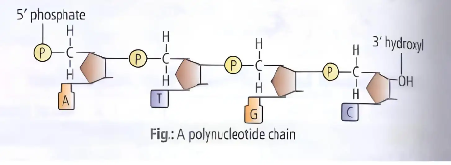
Structure of a Polynucleotide Chain
| Class | Bases |
|---|---|
| Purines | Adenine (A), guanine (G) |
| Pyrimidines | Cytosine (C), thymine (T), uracil (U) |
From Base to Polynucleotide
- Nitrogenous base attaches to the sugar's carbon by an N-glycosidic linkage → nucleoside.
- Phosphate attaches to the nucleoside's hydroxyl group → nucleotide.
- The phosphate links the carbon of one sugar to the carbon of the next → polynucleotide chain.
Nucleosides and Linkages
- Nucleoside examples: adenosine, guanosine, cytidine and uridine; DNA forms include deoxyadenosine, deoxyguanosine, deoxycytidine and deoxythymidine.
- Phosphodiester linkage: joins nucleotides; successive nucleotides form a chain with distinct and ends.
- RNA feature: its ribose has an extra -OH group; thymine is also known as 5-methyl uracil.
Identification of DNA
- 1869Friedrich Miescher identified an acidic substance in the nucleus and named it nuclein.
Structure of DNA
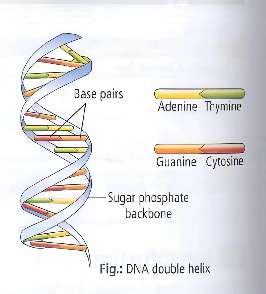
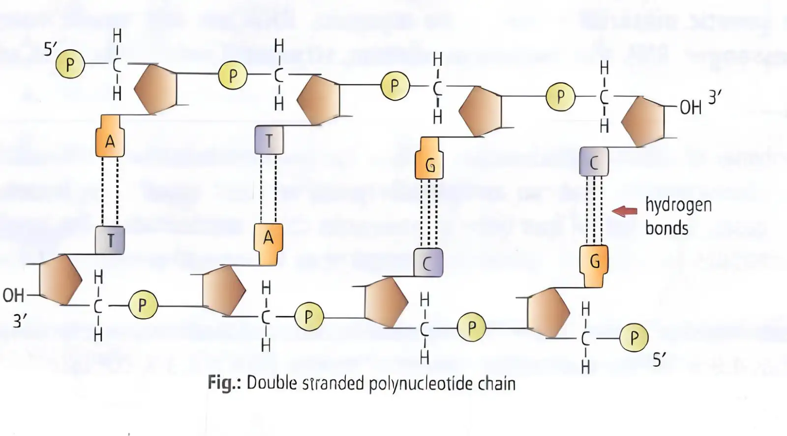
In double-stranded DNA, purines equal pyrimidines; adenine pairs with thymine and guanine with cytosine. The combined A–T and G–C proportions need not be equal.
Chargaff's Species-Specific Ratio
- A + T to G + C ratio: may differ between species but remains constant within a given species.
| Feature | Description |
|---|---|
| Strands and backbone | Two polynucleotide chains; sugar–phosphate backbones lie outside and bases project inward. |
| Polarity | Strands are antiparallel: one runs , the other . |
| Base pairing | A pairs with T by two hydrogen bonds; G pairs with C by three. Each pair contains one purine and one pyrimidine. |
| Helix | The two chains coil into a right-handed helix. |
| Dimensions | Pitch is 3.4 nm with about 10 bp per turn; adjacent base pairs are about 0.34 nm apart. |
| Stability | Stacking of successive base-pair planes, together with hydrogen bonding, stabilises the helix. |
Central Dogma
Information Flow
- DNA → RNA → protein (central dogma).
- In some viruses, information flows from RNA to DNA by reverse transcription.


Packaging of the DNA Helix
Use the distance between adjacent base pairs, approximately 0.34 nm.
DNA Packaging in Prokaryotes
- DNA packaging: Long DNA molecules are compacted so they can fit within the small cell.
- Prokaryotes: DNA is supercoiled with the help of RNA and basic non-histone proteins such as polyamines, forming the nucleoid.
DNA Packaging in Eukaryotes
Eukaryotic DNA Packing
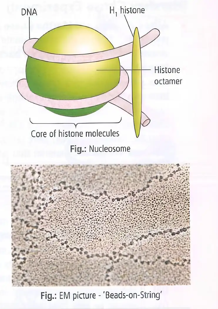
- DNA wraps around histone octamers → nucleosomes.
- Nucleosomes form beads-on-a-string chromatin → chromatin fibres.
- Fibres coil and condense at metaphase → chromosomes.
| Point | Euchromatin | Heterochromatin |
|---|---|---|
| Packing | Loosely packed | Densely packed |
| Staining | Light staining | Dark staining |
| Transcription | Transcriptionally active | Transcriptionally inactive |
Linker DNA and Chromatosome
- Linker (interbead) DNA: DNA connecting adjacent nucleosomes; it is associated with H₁, also called the plugging or marker protein.
- Chromatosome: a nucleosome together with its linker DNA.
State the base-pairing rules and the number of hydrogen bonds in each pair.
What is a nucleosome, and what is its usual DNA length?
Quick recap
- DNA is generally a double-stranded polymer of deoxyribonucleotides; RNA differs in sugar and uses uracil instead of thymine.
- Chargaff's rules are , and ; the A+T to G+C ratio varies by species.
- DNA strands are antiparallel and right-handed, with A–T and G–C pairing and base stacking contributing to stability.
- Genetic information usually flows DNA → RNA → protein; some viruses use reverse transcription from RNA to DNA.
- Prokaryotic DNA is supercoiled into a nucleoid; eukaryotic DNA wraps around histone octamers to form nucleosomes and chromatin.
- Euchromatin is loosely packed and active; heterochromatin is densely packed and inactive.
The Search for Genetic Material
Evidence for Genetic Material
- Evidence: Both indirect and direct experiments established that DNA is the genetic material in most organisms.
Griffith’s Transforming Principle
Transformation — A change in bacterial traits caused by uptake of genetic material from another source.
Griffith’s Experiment (1928)
- Organism: Frederick Griffith studied *Streptococcus pneumoniae*, which has virulent S and non-virulent R strains.
- S strain: Forms smooth colonies and is surrounded by a mucous polysaccharide sheath; it causes pneumonia.
- R strain: Forms irregular, rough colonies and is non-virulent.
- Finding: A factor from heat-killed S bacteria transformed live R bacteria into virulent, smooth-coated bacteria.
| Injection | Result |
|---|---|
| Live S bacteria | Mice die |
| Live R bacteria | Mice live |
| Heat-killed S bacteria | Mice live |
| Heat-killed S + live R bacteria | Mice die; live R bacteria are transformed into virulent S-type bacteria |
Interpretation
- Heat-killed S cells provide a transforming principle.
- Live R cells take up this principle and gain the ability to form a smooth polysaccharide coat.
- Transformed R cells become virulent S-type bacteria.
Identifying the Transforming Principle
Avery–MacLeod–McCarty Experiment (1933–44)
- Researchers: Oswald Avery, Colin MacLeod and Maclyn McCarty tested which molecule from heat-killed S cells transformed live R cells.
- DNA test: Purified DNA from S bacteria caused R bacteria to transform into S-type cells.
- Protein and RNA tests: Proteases and RNases did not stop transformation, so protein and RNA were not the transforming substance.
- Conclusion: DNase inhibited transformation; DNA is the hereditary material responsible for transformation.
| Treatment | Effect on transformation | Inference |
|---|---|---|
| Protease | Transformation continued | Protein is not the transforming principle |
| RNase | Transformation continued | RNA is not the transforming principle |
| DNase | Transformation was inhibited | DNA is the transforming principle |
Hershey–Chase Experiment
DNA Is the Genetic Material (1952)
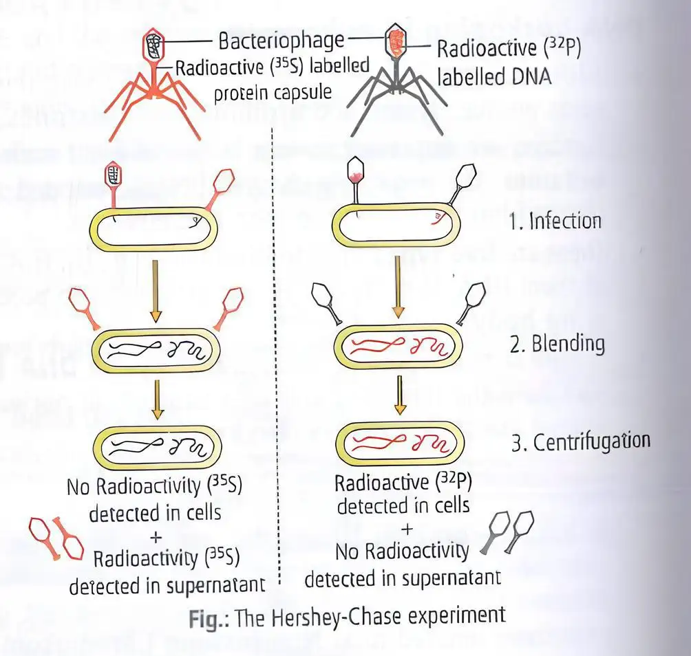
- Researchers: Alfred Hershey and Martha Chase used bacteriophages, viruses that infect bacteria, to test whether DNA or protein carries genetic information.
- DNA label: Phage DNA was labelled with radioactive phosphorus, .
- Protein label: Phage protein was labelled with radioactive sulphur, .
- Result: Bacteria infected with phages containing radioactive DNA became radioactive, showing that phage DNA entered the bacterial cells.
- Conclusion: DNA, not protein, enters bacterial cells and carries genetic information.
Properties of Genetic Material
Essential Criteria
- Replication: Must be able to generate its own replica.
- Stability: Must remain chemically and structurally stable enough to preserve information.
- Mutation: Must allow slow changes that provide variation required for evolution.
- Expression: Must be able to express itself as Mendelian characters.
| Point | DNA | RNA |
|---|---|---|
| Replication | Can direct its own duplication through complementary base pairing. | Can also direct its own duplication through complementary base pairing. |
| Chemical and structural stability | Less reactive and more stable; complementary DNA strands can reassociate under suitable conditions after heating separates them. | The reactive group makes RNA labile and easily degradable; RNA is also catalytic and reactive. |
| Mutation | Can mutate, generally more slowly than RNA. | Can mutate faster because it is less stable; RNA viruses with short life spans can evolve rapidly. |
| Expression | Depends on RNA for protein synthesis. | Can directly code for protein synthesis; the protein-synthesis machinery evolved around RNA. |
| Base associated with stability | Thymine in place of uracil contributes additional stability. | Contains uracil in place of thymine. |
| Typical role | Better suited for storage of genetic information. | Better suited for transmission of genetic information. |
Which Nucleic Acid Is the Better Genetic Material?
- Both: DNA and RNA can direct replication, mutate and express genetic information.
- DNA advantage: Greater chemical and structural stability makes DNA better for long-term storage of information.
- RNA exception: Some viruses use RNA as genetic material, including Tobacco Mosaic virus and Qβ bacteriophage.
- RNA-virus consequence: RNA’s instability can cause faster mutation; short-lived RNA viruses can evolve rapidly.
What did Griffith’s experiment show?
How did Avery, MacLeod and McCarty identify DNA as the transforming principle?
Quick recap
- Griffith found that heat-killed S bacteria could transform live R bacteria into virulent S-type cells.
- Avery, MacLeod and McCarty showed that DNase inhibited transformation, identifying DNA as the transforming principle.
- Hershey and Chase used -labelled DNA and -labelled protein to show that phage DNA enters bacteria.
- Genetic material must replicate, remain stable, permit mutation and express characters.
- DNA is more stable than RNA and is generally better for storing genetic information.
- Some viruses use RNA as genetic material; its greater instability can promote faster mutation.
RNA World
RNA as the First Genetic Material
- RNA-first hypothesis: RNA is considered the first genetic material and could act both as genetic material and as a catalyst.
- RNA world: Essential processes such as metabolism, translation and splicing are thought to have evolved around RNA; its catalytic reactivity also made it unstable.
Evolution of DNA
Why DNA is More Stable
- Origin: DNA evolved from RNA through chemical modifications that made it more stable.
- Double-stranded structure: A complementary strand helps DNA resist changes.
- Repair: DNA can use its complementary strand in a process of repair, further reducing changes.
| Point | RNA | DNA |
|---|---|---|
| Role / origin | First genetic material; also acted as a catalyst | Evolved from RNA after chemical modifications |
| Stability | Reactive and unstable | More stable; double-stranded structure and repair help resist changes |
Main Forms of RNA
Non-genetic RNA
- Classification: The three main forms are messenger RNA (mRNA), transfer RNA (tRNA) and ribosomal RNA (rRNA), classified by molecular size and function.
- Relative abundance: mRNA forms about 3.5% of cellular RNA, tRNA about 15% and rRNA about 80%.
| Type | Other names | Approximate share | Main feature / function |
|---|---|---|---|
| mRNA | Messenger, informational or template RNA | 3.5% | Transcribed from a gene and translated by ribosomes to produce protein. |
| tRNA | Transfer, soluble, acceptor or adapter RNA | 15% | Has an amino-acid-binding site opposite its anticodon site. |
| rRNA | Ribosomal RNA | 80% | A component of ribosomes, the cell's protein-synthesising factories; formed in the nucleolus. |
mRNA and Protein Synthesis
- Gene is transcribed into mRNA
- mRNA is translated by ribosomes
- Protein is produced
Why is DNA more stable than RNA?
Quick recap
- RNA is considered the first genetic material and could act as both genetic material and catalyst.
- RNA is reactive and unstable; DNA evolved from RNA with stabilising chemical modifications.
- DNA's complementary strand and repair process help protect it from changes.
- The three main RNA forms are mRNA, tRNA and rRNA.
- Their approximate shares of cellular RNA are 3.5%, 15% and 80%, respectively.
- mRNA carries information for protein production, tRNA binds amino acids, and rRNA forms part of ribosomes.
Replication
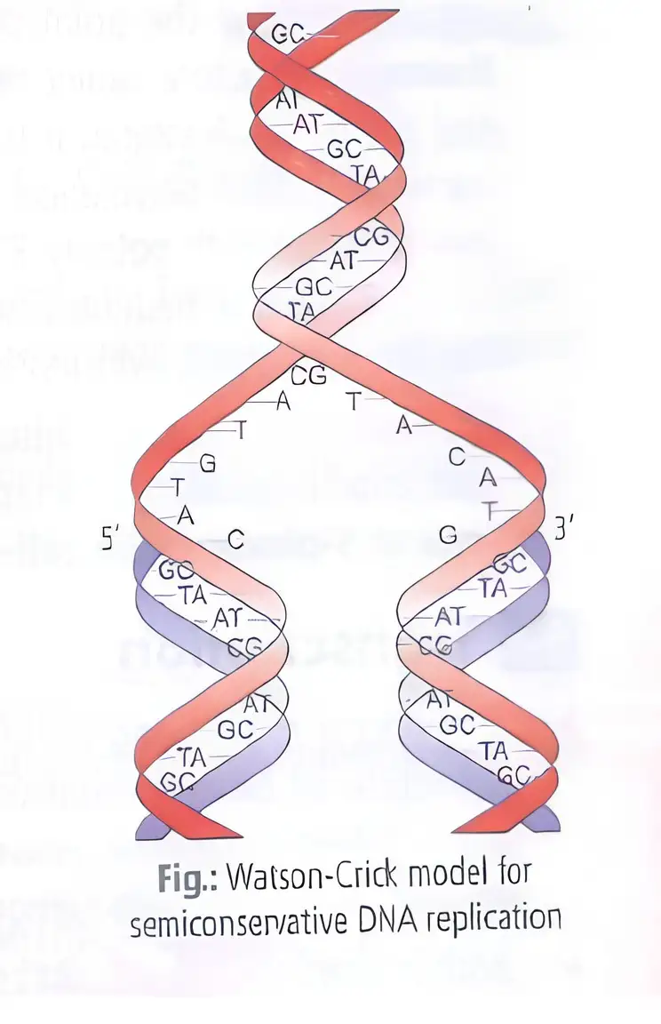
DNA Replication and the Semiconservative Model
Experimental Proof
Meselson and Stahl experiment
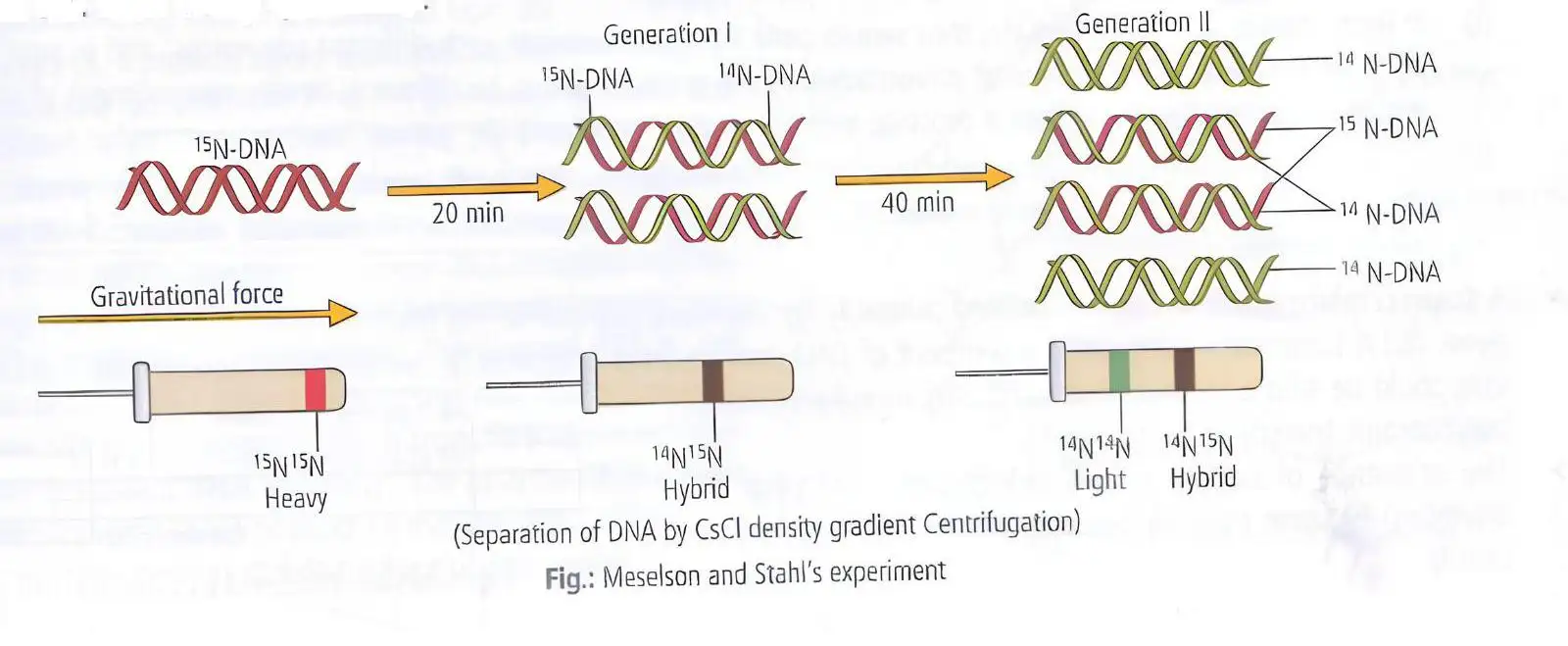
- Researchers and date: Matthew Meselson and Franklin Stahl demonstrated semiconservative replication in *E. coli* in 1958.
- Method and result: They labelled bacterial DNA with heavy nitrogen, , then transferred the cells to a medium. DNA from the generation had intermediate density.
- Conclusion: The intermediate-density DNA showed that each new DNA molecule contained one parental strand and one newly made strand.
Confirmation in plant chromosomes
- Taylor and colleagues (1958): Used radioactive thymidine to track newly synthesised DNA in chromosomes of *Vicia faba* (faba bean).
- Conclusion: Chromosomal DNA also replicates semiconservatively.
Replication Machinery and Enzymes
Energy, speed and accuracy
- DNA-dependent DNA polymerase: Uses a DNA template to polymerise deoxyribonucleotides; it must work rapidly and efficiently.
- Energy source: Replication requires abundant energy from the breakdown of deoxyribonucleotide triphosphates.
- E. coli rate: Its genome contains about bp and is replicated in about 18 minutes, at an average rate of approximately 2,000 bp per second.
- Scale comparison: The human diploid genome contains about bp.
- Errors: Mistakes made during replication can result in mutations.
| Component | Role |
|---|---|
| Helicase | Unwinds and separates the DNA strands at the origin. |
| Single-stranded binding proteins (ssBPs) | Stabilise the separated DNA strands. |
| Topoisomerases | Relieve tension from supercoiling by nicking and resealing a DNA strand. |
| RNA primer | A short RNA strand synthesised at the end of a new DNA strand; it provides the starting point needed by DNA polymerase. |
| DNA polymerase | Adds nucleotides to synthesise DNA only in the direction. |
| DNA ligase | Joins the fragments produced during discontinuous synthesis. |
Unwinding and formation of the replication fork
- Helicase acts at the origin of replication and separates the strands.
- ssBPs stabilise the exposed strands.
- Unwinding creates supercoiling and tension in the still-coiled DNA.
- Topoisomerases relieve the tension by temporarily nicking and then resealing a strand.
- DNA opens progressively in both directions, forming Y-shaped replication forks.
Priming and strand synthesis
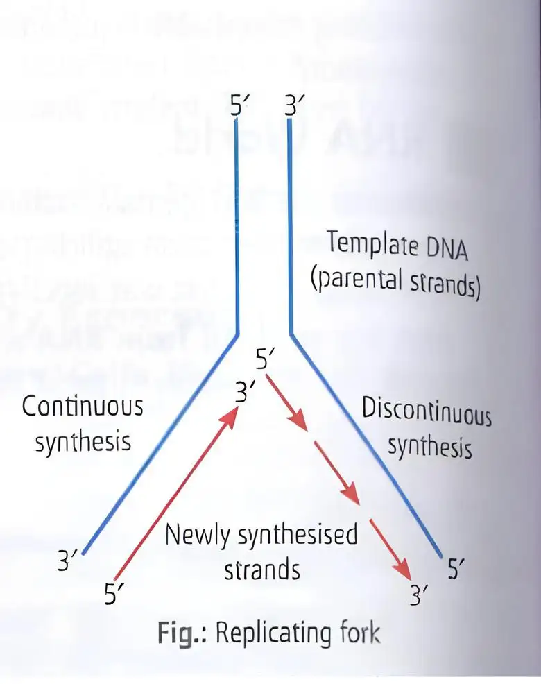
- RNA primer: A short RNA strand is synthesised at the end of a new DNA strand; DNA polymerase needs it to initiate a new DNA chain.
- Direction: DNA polymerase adds nucleotides only in the direction.
- Continuous synthesis: Occurs on the template with polarity.
- Discontinuous synthesis: Occurs on the template with polarity because only a short stretch is exposed at a time; DNA ligase later joins the fragments.
| Point | Continuous synthesis | Discontinuous synthesis |
|---|---|---|
| Template polarity | ||
| Reason | The template polarity allows synthesis to proceed continuously. | Only a short stretch of template is exposed at a time, so synthesis occurs in fragments. |
| Joining | No fragment-joining step is required. | DNA ligase joins the separately synthesised fragments. |
Initiation of Replication
- Origin of replication: Replication begins at a specific DNA region, not at a random point; *E. coli* DNA has a definite origin.
- Eukaryotes: DNA replication takes place during the S phase of the cell cycle.
Why is DNA replication called semiconservative?
Why is synthesis discontinuous on one DNA template?
Quick recap
- DNA replication is semiconservative: each daughter molecule has one old and one new strand.
- Meselson and Stahl used -labelled *E. coli* DNA and observed intermediate-density DNA.
- Taylor and colleagues supported semiconservative replication in *Vicia faba* chromosomes using radioactive thymidine.
- Helicase unwinds DNA; ssBPs stabilise strands; topoisomerases relieve supercoiling.
- An RNA primer is synthesised at the end of a new DNA strand; DNA polymerase extends DNA only in the direction.
- Synthesis is continuous on one template and discontinuous on the other; DNA ligase joins the fragments.
- Replication starts at an origin; eukaryotic DNA replication occurs in S phase.
Transcription
Meaning and Scope of Transcription
Why only one DNA strand is transcribed
- Different messages: If both strands were templates, they would produce RNAs with different sequences and could specify different proteins from the same DNA region.
- Double-stranded RNA: The two complementary RNAs could pair with each other, preventing translation into protein.
Transcription Unit and DNA Strands
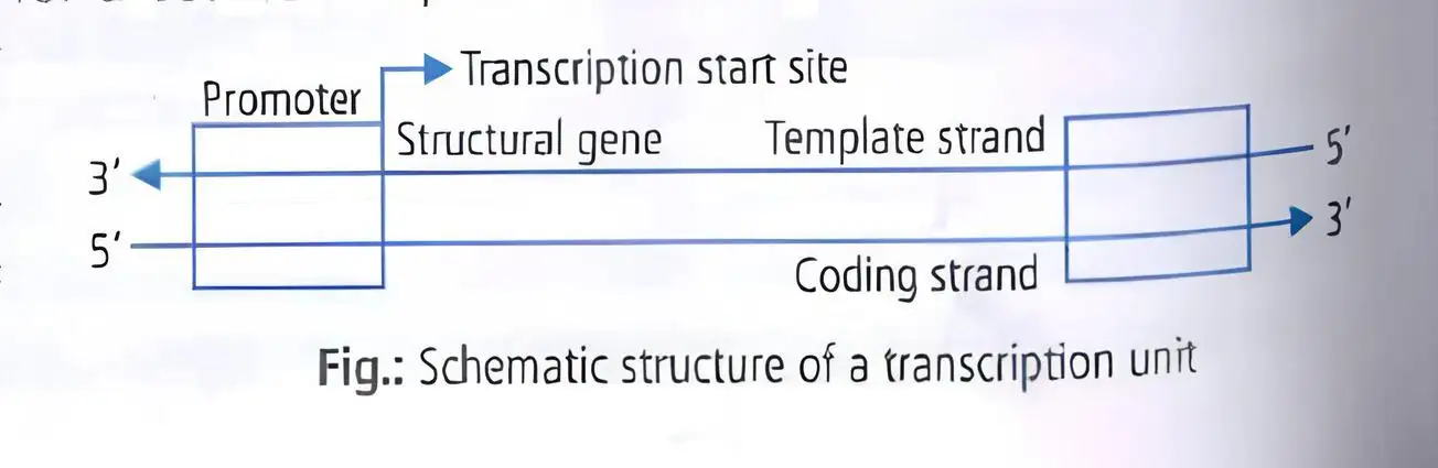
Transcription unit — A DNA region consisting of a promoter, a structural gene and a terminator.
- Promoter: DNA region where RNA polymerase binds to begin transcription.
- Structural gene: DNA sequence that is transcribed into RNA; it may be monocistronic or polycistronic.
- Terminator: DNA region that signals the end of transcription.
Cistron — A segment of DNA that codes for a polypeptide.
| Type | Typical occurrence |
|---|---|
| Monocistronic | Mostly eukaryotes |
| Polycistronic | Mostly prokaryotes |
Template and coding strands
- Template strand: The strand read by RNA polymerase to make RNA.
- Coding strand: Its sequence matches the RNA sequence, except DNA has thymine where RNA has uracil.
Bacterial RNA Types and Transcription
| RNA | Role in protein synthesis |
|---|---|
| mRNA | Provides the template for protein synthesis. |
| tRNA | Brings amino acids and reads the genetic code. |
| rRNA | Has structural and catalytic roles during translation. |
RNA polymerase
- Enzyme: Transcription requires DNA-dependent RNA polymerase.
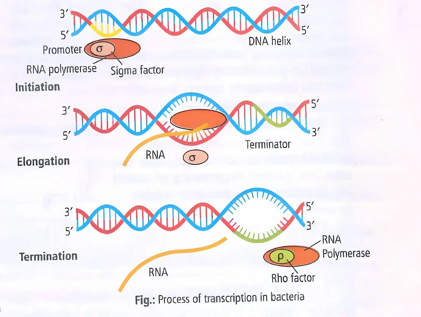
- Holoenzyme: The complete bacterial enzyme consists of a core enzyme and a sigma factor.
- Sigma factor: Recognises the DNA start signal and directs the enzyme to the promoter, increasing transcription specificity.
| Stage | Key events |
|---|---|
| Initiation | Holoenzyme recognises the promoter near the region and binds it. Binding near the region tightens, locally untwisting DNA. |
| Elongation | After about – nucleotides are polymerised, sigma dissociates. RNA is extended in the direction as polymerase untwists DNA ahead. |
| Termination | Rho factor follows polymerase on the RNA. At a pause site, rho catches up and unwinds the DNA–RNA hybrid, releasing the polymerase. |
Elongation details
- Transcription bubble: A single-stranded DNA bubble of about travels with the enzyme.
- RNA–DNA pairing: About newly made RNA bases remain paired with the single-stranded DNA in the bubble; the rest of the RNA exits as a single strand.
Bacterial transcription and translation
- Location: Both transcription and translation occur in the cytoplasm.
- mRNA processing: Bacterial mRNA requires no processing to become active.
Transcription in Eukaryotes
| Polymerase | Main RNA products |
|---|---|
| RNA polymerase I | 28S, 18S and 5.8S rRNAs |
| RNA polymerase II | hnRNA (pre-mRNA), the precursor of mRNA |
| RNA polymerase III | tRNA, 5S rRNA and snRNA |
Location and mRNA maturation
- Location: Transcription occurs in the nucleus; processed mRNA moves to the cytoplasm for translation.
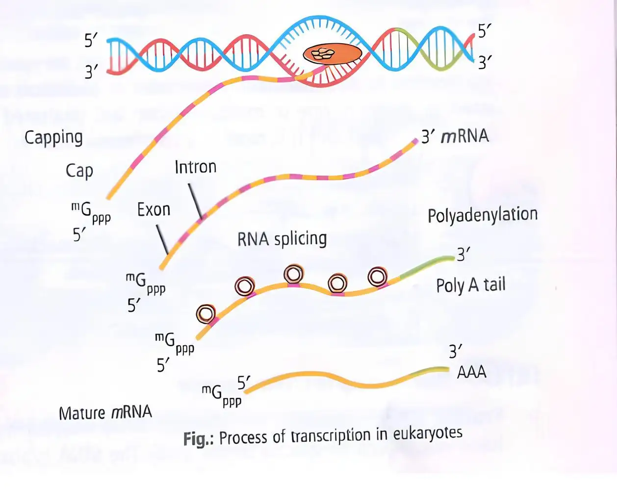
- Primary transcript: The longer nuclear hnRNA (pre-mRNA) contains coding exons and non-coding introns.
- Maturation: Processing of the primary RNA transcript produces mature mRNA.
| Process | What happens |
|---|---|
| Capping | A modified guanosine triphosphate cap, 7-methylguanosine (7mG), is added at the end. |
| Splicing | Introns are removed; adjacent exons are joined by ligase to form the continuous coding sequence. |
| Polyadenylation | A poly-A tail of about – residues is added at the end. |
Key features of RNA processing
- Template-independent additions: The cap and poly-A tail are added without using a template.
- RNA world: Splicing is considered evidence for the dominance of the RNA world.
Why is only one DNA strand transcribed?
What are the products of eukaryotic RNA polymerases I, II and III?
Quick recap
- Transcription copies one DNA strand from a selected segment into RNA; RNA uses uracil instead of thymine.
- A transcription unit contains a promoter, structural gene and terminator; structural genes may be monocistronic or polycistronic.
- Bacterial transcription uses a core enzyme and sigma factor, and proceeds through initiation, elongation and rho-assisted termination.
- Bacteria transcribe and translate in the cytoplasm, and their mRNA needs no processing.
- Eukaryotic polymerases I, II and III transcribe distinct RNA groups in the nucleus.
- Eukaryotic pre-mRNA is capped, spliced and given a poly-A tail before translation.
Genetic Code
Meaning and Triplet Nature of the Genetic Code
Deciphering the Genetic Code
- Har Gobind Khorana developed chemical methods to synthesise RNA with defined base sequences, including homopolymers and copolymers.
- Severo Ochoa’s enzyme polynucleotide phosphorylase helped synthesise RNA with defined sequences in a template-independent manner.
- First codon identified: Nirenberg and Matthaei identified , which codes for phenylalanine.
Characteristics of the Genetic Code
Do not interpret an intermediate as coding for valine: in the standard genetic code, codes for methionine. Valine is coded by , , and .
Wobble Hypothesis
- Proposed by: F. H. C. Crick in 1965.
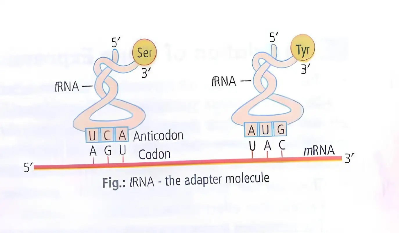
- Wobble: The third codon base pairs less strictly with the anticodon; the first two bases mainly specify the codon.
- Consequence: One tRNA can recognise multiple codons that differ only at the third base.
Recognition of the Genetic Code
- Nobel Prize, 1968: Holley, Nirenberg and Khorana received the prize for deciphering the genetic code.
| Amino acid or signal | Codons |
|---|---|
| Phenylalanine (Phe) | , |
| Leucine (Leu) | , , , , , |
| Serine (Ser) | , , , , , |
| Tyrosine (Tyr) | , |
| Cysteine (Cys) | , |
| Tryptophan (Trp) | |
| Proline (Pro) | , , , |
| Histidine (His) | , |
| Glutamine (Gln) | , |
| Arginine (Arg) | , , , , , |
| Isoleucine (Ile) | , , |
| Methionine (Met) | |
| Threonine (Thr) | , , , |
| Asparagine (Asn) | , |
| Lysine (Lys) | , |
| Valine (Val) | , , , |
| Alanine (Ala) | , , , |
| Aspartic acid (Asp) | , |
| Glutamic acid (Glu) | , |
| Glycine (Gly) | , , , |
| Stop | , , |
Mutations and the Genetic Code
- Point mutation: A single base-pair change in the beta-globin gene replaces glutamate with valine, causing sickle-cell anaemia.
- Frameshift mutation: Insertion or deletion of one or two bases shifts the reading frame; changes in multiples of three do not shift it.
- Other mutations: Nonsense mutations create a stop codon, missense mutations change an amino acid, and silent mutations change a codon without changing its amino acid.
tRNA: The Adapter Molecule
What is the wobble hypothesis?
How do insertions or deletions affect the reading frame, and what does this show about codons?
Quick recap
- A codon is a triplet; four RNA bases produce 64 possible codons.
- The genetic code relates DNA base sequences to polypeptide amino-acid sequences through RNA codons.
- The code is universal, degenerate, non-overlapping and unambiguous.
- codes for methionine; , and are stop codons.
- Wobble at the third base lets one tRNA recognise multiple codons.
- Insertions or deletions not in multiples of three cause frameshifts; this supports contiguous triplet reading.
- tRNA pairs its anticodon with an mRNA codon and carries the corresponding amino acid.
Translation
Translation and Amino Acid Activation
Translation — The polymerisation of amino acids into a polypeptide, directed by the sequence of bases in mRNA.
Amino Acid Activation
- Amino acid sequence: The order of amino acids in the polypeptide is determined by the codon sequence in mRNA.
- Charging of tRNA: Each amino acid is activated using ATP and linked to its cognate tRNA; this is called aminoacylation or tRNA charging.
- Peptide bond: Amino acids are joined by peptide bonds, and their formation requires energy.
Ribosomes and the Translational Unit
Ribosome
- Function: Ribosomes are the cellular machinery for protein synthesis and contain structural RNAs and about 80 different proteins.
- Subunits: An inactive ribosome consists of a large and a small subunit; translation begins when the small subunit encounters mRNA.
- Binding sites: The large subunit has two sites for binding successive amino acid–tRNA complexes.
- Catalytic role: The ribosome catalyses peptide-bond formation; in bacteria, 23S rRNA acts as a ribozyme.
Translational unit — The mRNA sequence between the start codon AUG and a stop codon that codes for a polypeptide.
Untranslated Regions
- 5′ UTR: Lies before the start codon; 3′ UTR: lies after the stop codon.
- Role: UTRs are not translated but are required for efficient translation.
Stages of Translation
| Stage | Key events |
|---|---|
| Initiation | The ribosome binds mRNA at the start codon AUG, recognised by the initiator tRNA. |
| Elongation | Charged tRNAs pair their anticodons with complementary mRNA codons; the ribosome moves codon by codon and adds amino acids to the growing polypeptide. |
| Termination | A release factor binds the stop codon, ending translation and releasing the completed polypeptide from the ribosome. |
Protein Biosynthesis
Post-Translational Modifications
- Maturation: Newly synthesised proteins undergo post-translational modifications (PTMs) to become mature functional products.
- Specificity: PTMs are mostly enzyme-catalysed; enzymes recognise specific target sequences in particular proteins.
Polyribosomes
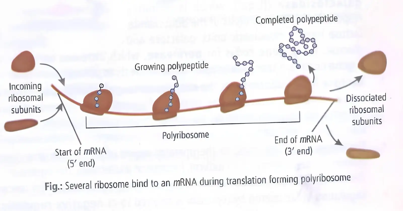
Polyribosome — A group of several ribosomes translating the same mRNA at the same time.
- Formation: After the first ribosome moves far enough beyond the start codon, another ribosome can attach to the same mRNA; further ribosomes may join.
- Result: Multiple ribosomes produce many copies of the same polypeptide simultaneously.
- Occurrence: Polyribosomes are found in both prokaryotes and eukaryotes.
What is tRNA charging?
How does translation terminate?
Quick recap
- Translation uses the mRNA codon sequence to assemble amino acids into a polypeptide.
- ATP-dependent amino acid activation and tRNA charging prepare amino acids for translation.
- Ribosomes contain structural RNAs and proteins; bacterial 23S rRNA catalyses peptide-bond formation.
- Translation proceeds through initiation at AUG, codon-by-codon elongation and release-factor-mediated termination.
- Post-translational modifications help convert newly made proteins into mature products.
- Polyribosomes let multiple ribosomes translate one mRNA simultaneously in both prokaryotes and eukaryotes.
Regulation of Gene Expression
Gene expression — Gene expression produces functional products; metabolic, physiological and environmental conditions regulate when genes are expressed.
Need-based gene expression
- Example: E. coli uses beta-galactosidase to hydrolyse lactose into glucose and galactose, which can be used for energy.
- Need-based expression: Without lactose, the bacterium does not need to synthesise beta-galactosidase.
Types of Gene Regulation
| Type | How it works | Alternative term |
|---|---|---|
| Negative regulation | Genes express until their activity is suppressed by a product of a regulatory gene. | Repressible regulation |
| Positive regulation | Genes remain non-expressed unless induced. | Inducible regulation |
Levels of Gene Regulation
| Organism group | Main control points |
|---|---|
| Eukaryotes | Transcription; RNA processing, including splicing; mRNA transport from nucleus to cytoplasm; translation |
| Prokaryotes | Predominantly control the initiation of transcription |
Prokaryotic transcriptional control
- RNA polymerase: Accessory proteins regulate its activity at a promoter.
- Operator: A DNA sequence, usually next to the promoter in an operon, whose protein interactions regulate promoter accessibility.
- Specificity: Each operon has a specific operator and repressor; the lac repressor interacts specifically with the lac operator.
The Lac Operon
Operon — A bacterial arrangement in which a polycistronic structural gene is regulated by a common promoter and regulatory genes.
Operon examples and discovery
- Examples: lac, trp, ara, his and val operons.
- First elucidated: Geneticist Francois Jacob and biochemist Jacques Monod first described the lac operon system.
| Gene | Product | Function |
|---|---|---|
| i | Repressor | Regulatory gene; i comes from “inhibitor”. The repressor is synthesised constitutively. |
| z | Beta-galactosidase | Hydrolyses lactose into glucose and galactose. |
| y | Permease | Increases cell permeability to beta-galactosides. |
| a | Transacetylase | Encodes the third structural-gene product involved in lactose metabolism. |
Induction and transcription
- Inducer: Lactose, or allolactose, acts as the inducer and switches the operon on.
- Lactose entry: When lactose is supplied and a preferred carbon source such as glucose is absent, permease transports lactose into the cell.
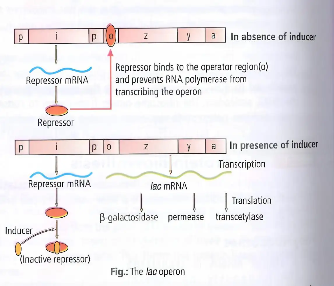
- Without inducer: The repressor binds the operator and prevents RNA polymerase from transcribing the operon.
- With inducer: Lactose or allolactose interacts with and inactivates the repressor, allowing RNA polymerase access to the promoter and transcription to proceed.
- Products: The z, y and a gene products are required for lactose metabolism.
Lac operon switching
- No inducer → repressor binds operator → transcription is blocked
- Lactose or allolactose present → repressor is inactivated → RNA polymerase transcribes the operon
- Transcription → beta-galactosidase, permease and transacetylase are produced
The lac operon is induced by lactose or allolactose, but the repressor-mediated control is negative regulation.
What happens to lac operon transcription when the inducer is absent?
Name the lac operon genes and their products.
Quick recap
- Gene expression is regulated by metabolic, physiological and environmental conditions.
- Eukaryotic regulation can occur during transcription, RNA processing, mRNA transport and translation.
- Prokaryotic regulation occurs predominantly at transcription initiation, through proteins acting at promoters and operators.
- An operon places polycistronic structural genes under common regulatory control.
- The lac operon contains regulatory gene i and structural genes z, y and a.
- Lactose or allolactose inactivates the lac repressor, allowing transcription.
Human Genome Project
Human Genome Project: Overview
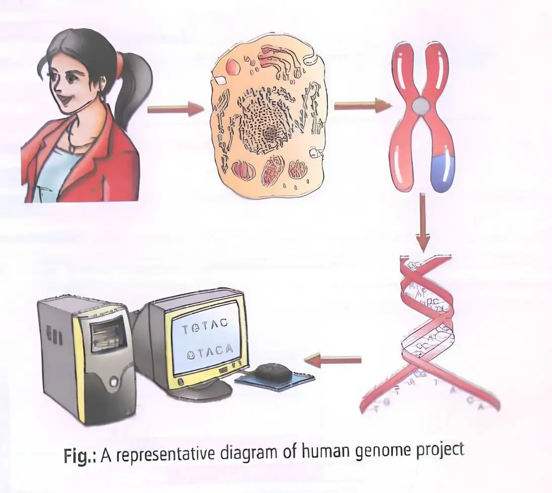
- Launch and completion: The Human Genome Project (HGP), an international mega-project, began in 1990 and was completed in 2003.
- Coordination: It was coordinated by the U.S. Department of Energy and the National Institutes of Health and helped drive the development of bioinformatics.
Goals of the HGP
- Genome analysis: Identify human genes and determine the sequence of the approximately 3 billion base pairs in human DNA.
- Resources and maps: Store and analyse the data, improve analysis tools, transfer technologies, develop genetic and physical maps, and address ethical, legal and social issues.
Why the Project Became Possible
- Genetic engineering: Techniques made it possible to isolate and clone any DNA segment.
- DNA sequencing: Simple, rapid sequencing techniques made large-scale genome analysis feasible.
Methodologies of the HGP
| Approach | Focus |
|---|---|
| Expressed Sequence Tags (ESTs) | Identify genes that are expressed as RNA. |
| Sequence annotation | Sequence the whole genome, including coding and non-coding regions, then assign functions to different regions. |
Sequencing and Assembly
- Sequencing: Automated DNA sequencers read fragments using the method developed by Frederick Sanger.
- Overlaps: Fragments were assembled by matching overlapping regions; this required generating overlapping DNA fragments.
- Computer analysis: The large number of sequences could not be aligned manually, so specialised computer programs were developed.
- Annotation: Sequences were annotated and assigned to chromosomes.
- Chromosome 1: Its sequence was completed in May 2006; it was the last of the 24 chromosome types sequenced (22 autosomes, X and Y).
Salient Features of the Human Genome
- Genome size and genes: The human genome has 3,164.7 million base pairs and about 30,000 genes; the average gene is about 3,000 bases, while the dystrophin gene is 2.4 million bases.
- Genome composition: About 99.9% of bases are the same among people, less than 2% of the genome codes for proteins, and repeated sequences make up a large portion.
- Variation and chromosomes: Chromosome 1 has the most genes (2,968) and the Y chromosome the fewest (231); about 1.4 million SNP locations were identified.
Applications of the HGP
What were the two main approaches used to sequence the human genome?
State four salient features revealed by the HGP.
Quick recap
- The HGP began in 1990 and was completed in 2003, coordinated by U.S. government agencies.
- Its goals included identifying genes, sequencing DNA, building maps, storing data and addressing ELSI.
- ESTs identify RNA-expressed genes; sequence annotation covers the whole genome and assigns functions.
- Automated Sanger-based sequencing, overlap assembly and computer programs supported genome analysis.
- The human genome contains about 3,164.7 million base pairs, with less than 2% coding for proteins.
- HGP data supports genome-wide research and understanding gene–protein interaction networks.
DNA Fingerprinting
DNA Fingerprinting and VNTRs
Polymorphism and Its Uses
- Polymorphism: Genetic variation arises through mutations; a DNA variant is considered polymorphic when it occurs in a population at a frequency greater than 0.01.
- Uses: DNA variation supports individual identification, forensic investigation and paternity testing, and helps study population and genetic diversity.
DNA Fingerprinting Technique
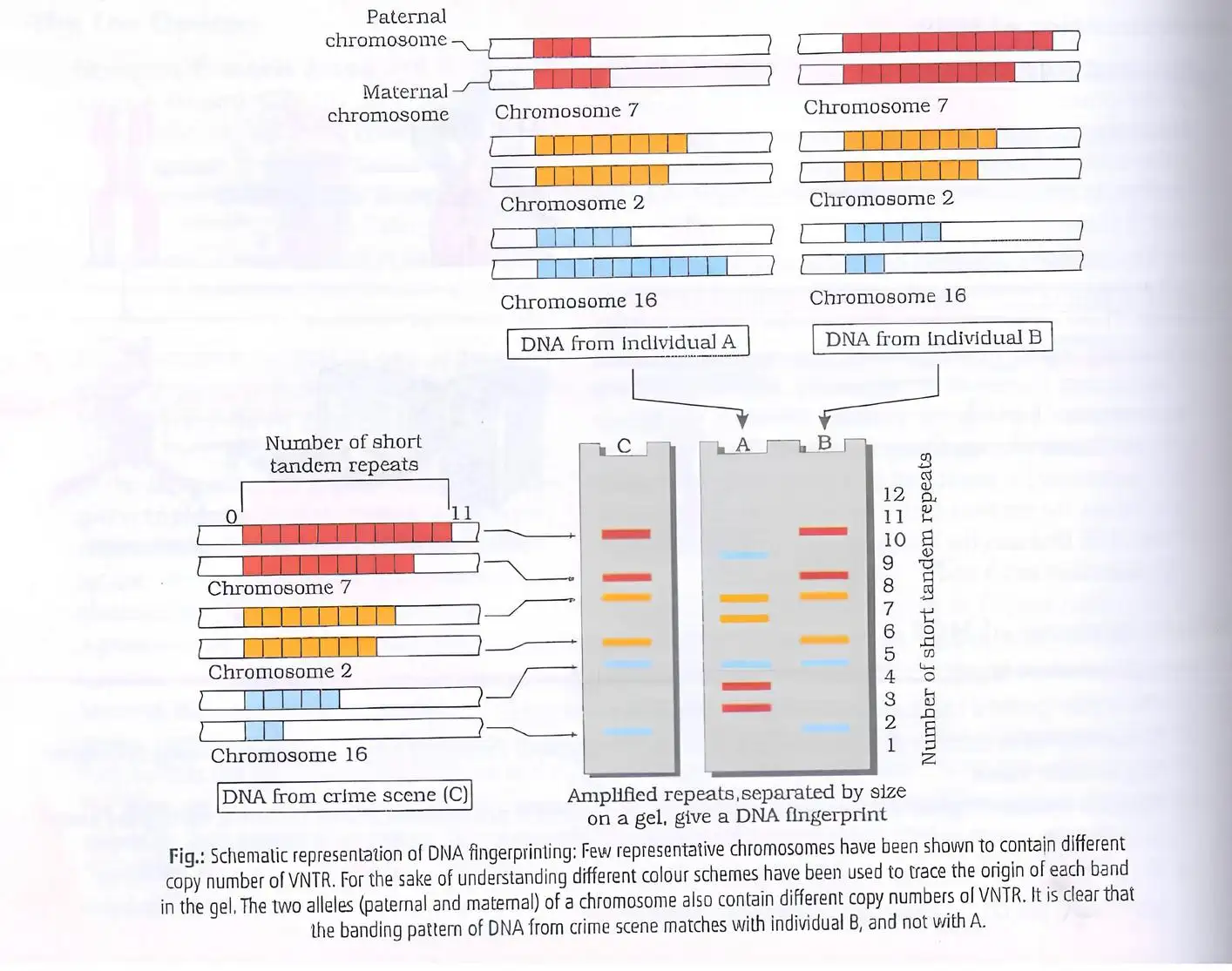
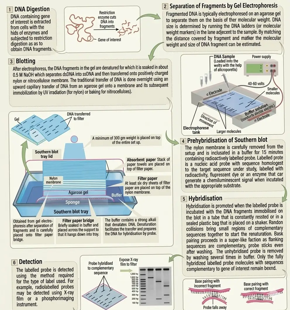
- Basis: Alec Jeffreys developed DNA fingerprinting using highly polymorphic repetitive DNA sequences called VNTRs.
- Pattern: DNA fragments are separated and detected using VNTR probes, producing a banding pattern characteristic of an individual, except in identical twins.
Southern Blotting
Applications of DNA Fingerprinting
- Forensic identification: DNA fingerprinting can help identify criminals and establish biological relationships, including paternity.
- Population studies: It is used to determine population and genetic diversity.
DNA Sequencing by Sanger’s Method
What are VNTRs, and why are they useful in DNA fingerprinting?
What are the main detection steps in the original DNA fingerprinting technique?
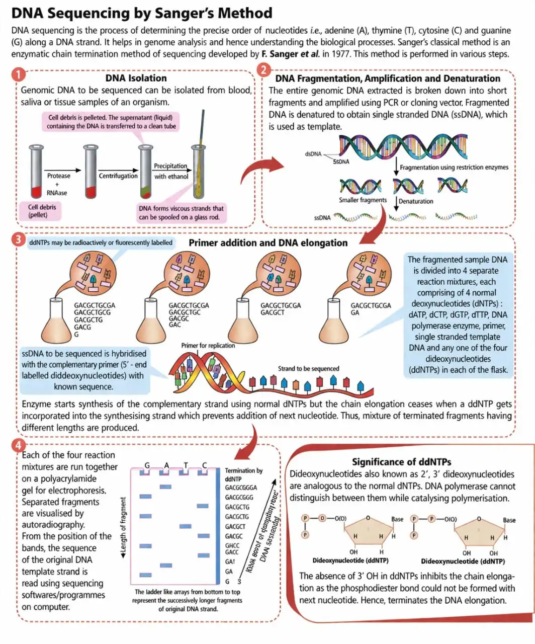
State the principle of Sanger’s DNA sequencing method.
Quick recap
- VNTRs are highly polymorphic repeated DNA regions used to identify individuals.
- Polymorphism results from mutation; DNA variants at a locus occur above a population frequency of 0.01.
- The original fingerprinting method uses restriction digestion, electrophoresis, Southern blotting, VNTR-probe hybridisation and autoradiography.
- Edwin Southern’s blotting method transfers DNA bands to a membrane for probe analysis.
- DNA fingerprinting is useful in forensics, biological relationship testing and studies of genetic diversity.
- Sanger sequencing is an enzymatic chain-termination method developed in 1977.
NEET Botany — ALL CHAPTERS
1
The Living World
2
Plant Kingdom
3
Morphology of Flowering Plants
4
Anatomy of Flowering Plants
5
The Unit of Life
6
Photosynthesis in Higher Plants
7
Respiration in Plants
8
Plant Growth and Development
9
Sexual Reproduction in Flowering Plants
10
Principles of Inheritance and Variation
11
Molecular Basis of Inheritance
12
Biotechnology :Principles and Processes
13
Biotechnology and its Applications