Class 12 PCB . Biology . Biology . Human Reproduction
Chapter 2: Human Reproduction
Ch 2
Biology
Class 12 PCB
The Male Reproductive System
Main Events in Human Reproduction
Overview
- Reproduction: Humans reproduce sexually and are viviparous; offspring develop inside the mother and are born alive.
- Gametogenesis: Formation of gametes—sperms in males and ova in females.
- Insemination: Transfer of sperms into the female genital tract; fertilisation is the fusion of male and female gametes to form a zygote.
- After fertilisation: The zygote develops into a blastocyst, which attaches to the uterine wall during implantation.
- Gestation and birth: Gestation is embryonic development; parturition is delivery of the baby. These reproductive events occur after puberty.
- Age-related difference: Sperm formation may continue in older men, whereas ovum formation ceases in women around age 50.
Organisation of the Male Reproductive System
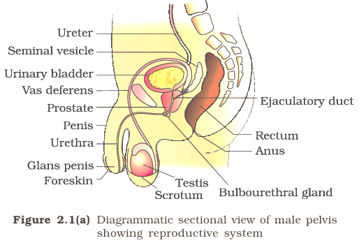
Male reproductive system — A pelvic system comprising a pair of testes, accessory ducts, accessory glands and external genitalia.
Relation to the Urinary System
- Connection: The reproductive system works closely with urinary-system structures such as the urethra and bladder, but the systems have distinct roles.
- Urethra: It carries urine from the bladder and provides the passage through the penis for sperm to leave the body.
Testes and Sperm Production
Testes and Seminiferous Tubules
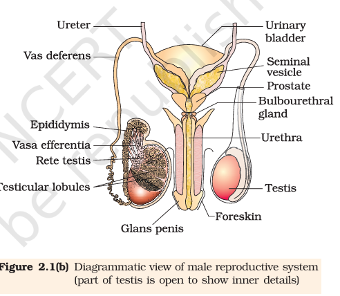
- Scrotum: The testes lie outside the abdominal cavity in the scrotum, which keeps them about – below internal body temperature—an essential condition for spermatogenesis.
- Size and structure: Each adult testis is oval, about – long and – wide, and has a dense covering.
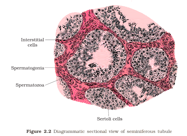
- Lobules: Each testis contains about 250 testicular lobules; each lobule contains 1–3 highly coiled seminiferous tubules.
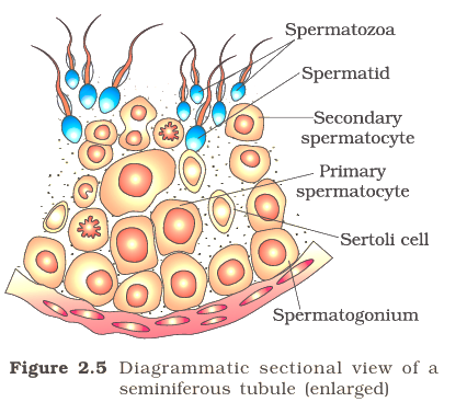
- Spermatogonia: Male germ cells lining the seminiferous tubules undergo meiotic divisions that ultimately lead to sperm formation.
- Sertoli cells: These tubule cells provide nutrition to developing germ cells.
Interstitial Spaces
- Contents: The spaces outside seminiferous tubules contain small blood vessels, Leydig cells and other immunologically competent cells.
- Leydig cells: These interstitial cells synthesise and secrete androgens, the testicular hormones.
Male Accessory Ducts
Male accessory ducts — The ducts that store and transport sperm from the testes to the outside through the urethra.
Route of Sperm
- Seminiferous tubules → rete testis → vasa efferentia.
- Vasa efferentia → epididymis, located along the posterior surface of each testis.
- Epididymis → vas deferens, which ascends into the abdomen and loops over the urinary bladder.
- The vas deferens receives a duct from the seminal vesicle and opens into the urethra as the ejaculatory duct.
- Urethra passes through the penis and opens outside at the urethral meatus.
Male Accessory Glands
Glands and Their Secretions
- Glands: Paired seminal vesicles, a prostate gland and paired bulbourethral glands.
- Seminal plasma: Secretions from these glands form seminal plasma, which is rich in fructose, calcium and certain enzymes.
- Bulbourethral glands: Their secretions also help lubricate the penis.
The Female Reproductive System
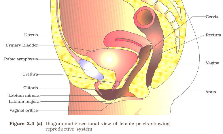
Female reproductive system — A pelvic system comprising a pair of ovaries, a pair of oviducts, the uterus, cervix, vagina and external genitalia.
Ovaries
- Functions: Ovaries produce ova and steroid hormones called ovarian hormones.
- Position and size: One ovary lies on each side of the lower abdomen; each is about – long and is connected to the pelvic wall and uterus by ligaments.
- Structure: A thin epithelium covers each ovary and encloses its stroma.
- Stroma: It has a peripheral cortex and an inner medulla.
Female Accessory Ducts
Oviducts
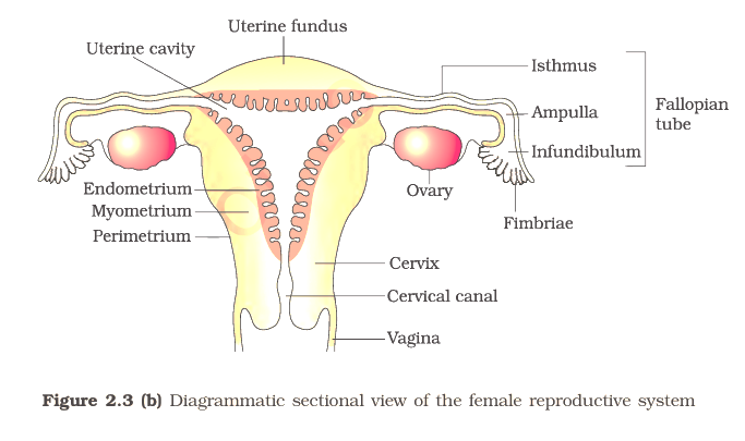
- Female accessory ducts: The oviducts (fallopian tubes), uterus and vagina form the accessory ducts.
- Length and route: Each fallopian tube is about – long and extends from near an ovary to the uterus.
- Infundibulum: The funnel-shaped part nearest the ovary; its finger-like fimbriae help collect the ovum after ovulation.
- Ampulla: The wider part of the oviduct following the infundibulum.
- Isthmus: The narrow-lumen final part that joins the uterus.
Uterus and Birth Canal
Uterine Structure and Functions
- Uterus: A single, inverted pear-shaped organ, also called the womb; ligaments connect it to the pelvic wall.
- Cervix: The uterus opens into the vagina through the narrow cervix. Its cavity is the cervical canal.
- Birth canal: The cervical canal and vagina together form the birth canal.
- Perimetrium: The thin, external membranous layer of the uterine wall.
- Myometrium: The thick middle layer of smooth muscle; it contracts strongly during delivery.
- Endometrium: The inner glandular layer lining the uterine cavity; it undergoes cyclical changes during the menstrual cycle.
Female External Genitalia and Mammary Glands
External Genitalia
- Mons pubis: A cushion of fatty tissue covered by skin and pubic hair.
- Labia majora: Fleshy folds extending down from the mons pubis and surrounding the vaginal opening.
- Labia minora: A pair of folds beneath the labia majora.
- Hymen: A membrane that often partially covers the vaginal opening. It may tear during intercourse, but can also break due to a fall or jolt, tampon insertion or some sports; in some women it remains intact after intercourse.
- Clitoris: A small, finger-like structure at the upper junction of the labia minora, above the urethral opening.
- Important: Hymen presence or absence is not a reliable indicator of virginity or sexual experience.
Mammary Glands
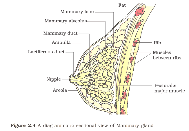
- Role in reproduction: A pair of mammary glands is structurally and functionally integrated with the reproductive system to support child care.
- Diagram structures: The mammary-gland section labels ducts, lobes, alveoli and the nipple.
Why are the testes located in the scrotum outside the abdominal cavity?
Name the male accessory ducts in the order followed by sperm, and state the functions of Sertoli and Leydig cells.
Name the three uterine layers and give one key feature of each.
Quick recap
- Reproductive sequence: Gametogenesis forms gametes; insemination transfers sperm; fertilisation forms a zygote, followed by blastocyst development, implantation, gestation and parturition.
- Male system: Testes, accessory ducts, accessory glands and external genitalia form the male reproductive system.
- Testes: Scrotal temperature supports spermatogenesis; seminiferous tubules form sperm, Sertoli cells nourish germ cells, and Leydig cells secrete androgens.
- Sperm route: Rete testis → vasa efferentia → epididymis → vas deferens → ejaculatory duct → urethra.
- Seminal plasma: Accessory-gland secretions are rich in fructose, calcium and certain enzymes; bulbourethral secretions help lubricate the penis.
- Female system: Ovaries produce ova and steroid hormones; oviducts, uterus, cervix, vagina and external genitalia complete the system.
- Key female structures: Fimbriae collect the ovum; endometrium changes cyclically; myometrium contracts during delivery; hymen status is not a reliable indicator of sexual experience.
Gametogenesis & Menstrual Cycle
Gametogenesis
- Gametogenesis: The process by which the primary sex organs produce gametes—sperms in males and ova in females.
- Meiosis: Gamete formation produces haploid cells, which combine during fertilisation to restore the diploid chromosome number.
Spermatogenesis and Its Hormonal Control
Hormonal Control of Spermatogenesis
- At puberty, increased hypothalamic GnRH stimulates the anterior pituitary.
- The pituitary releases LH and FSH.
- LH stimulates Leydig cells to produce androgens; androgens promote spermatogenesis.
- FSH acts on Sertoli cells, which release factors that support spermiogenesis.
Sperm Structure and Semen
Sperm Transport and Semen
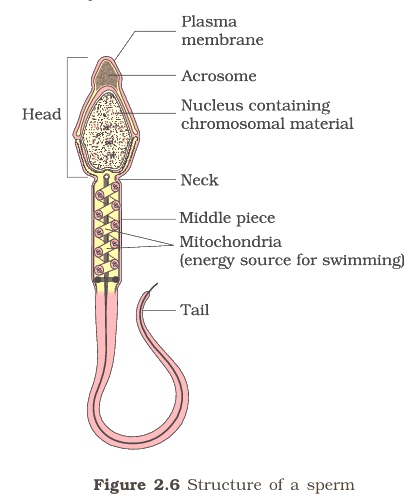
- Transport: Sperm move from the seminiferous tubules through the male accessory ducts.
- Maturation and motility: Secretions from the epididymis, vas deferens, seminal vesicles and prostate are essential.
- Semen: A mixture of sperm and seminal plasma.
- Hormonal support: Androgens maintain the functions of the male accessory ducts and glands.
Oogenesis
Follicle Development and Ovulation
- Primary follicle gains more granulosa-cell layers and a theca layer → secondary follicle.
- Secondary follicle develops into a tertiary follicle with a fluid-filled antrum; the theca forms inner theca interna and outer theca externa.
- The primary oocyte grows and completes meiosis I → a large haploid secondary oocyte and a small first polar body.
- The secondary oocyte retains most of the nutrient-rich cytoplasm; the first polar body's later fate is uncertain.
- Tertiary follicle matures into a Graafian follicle; the secondary oocyte develops a zona pellucida.
- The Graafian follicle ruptures and releases the secondary oocyte (often referred to as the ovum) → ovulation.
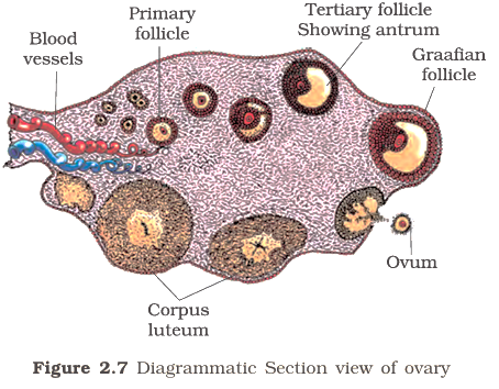
| Point | Spermatogenesis | Oogenesis |
|---|---|---|
| Begins | At puberty | During fetal development |
| Cell division | One primary spermatocyte forms four equal spermatids | One primary oocyte forms a large secondary oocyte and a small polar body |
| Cytoplasm | Divided relatively equally | Most retained in the secondary oocyte |
| Outcome | Four haploid sperm cells | One large haploid secondary oocyte and polar body |
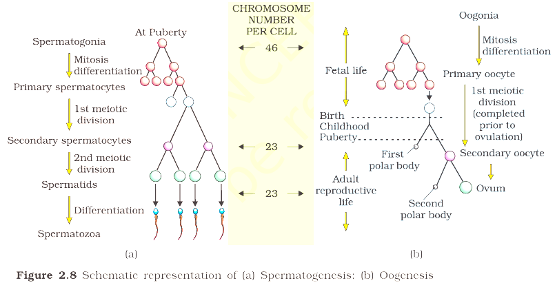
- Both processes produce haploid gametes with chromosomes; fertilisation restores the diploid number of .
- Unequal division in oogenesis preserves most of the primary oocyte's cytoplasm in the secondary oocyte.
Menstrual Cycle
| Phase | Main ovarian and uterine events | Hormonal changes / outcome |
|---|---|---|
| Menstrual phase (about 3–5 days) | The endometrial lining and its blood vessels break down; the material exits through the vagina. | Occurs when the released ovum is not fertilised; the cycle begins again. |
| Follicular phase | Ovarian follicles develop towards a mature Graafian follicle; the uterine endometrium regenerates and proliferates. | Pituitary LH and FSH gradually rise; growing follicles secrete oestrogens. |
| Ovulatory phase (around day 14) | The Graafian follicle ruptures and releases the secondary oocyte. | LH and FSH peak mid-cycle; the sharp LH surge triggers ovulation. |
| Luteal phase | The ruptured follicle's remains form the corpus luteum. | Corpus luteum secretes progesterone, which maintains the endometrium for possible implantation. |
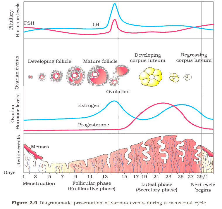
If Fertilisation Does Not Occur
- No fertilisation → corpus luteum degenerates.
- Corpus luteum degeneration → progesterone support falls.
- Endometrium disintegrates → menstruation begins and marks a new cycle.
Pregnancy and Menstrual Changes
- Endometrium: Its maintenance by progesterone is important for implantation and pregnancy.
- Pregnancy: Menstrual-cycle events stop, so menstruation does not occur.
- Missed menstruation: May indicate pregnancy, but stress, poor health and other causes can also disrupt menstruation.
Menstrual Hygiene
Trace spermatogenesis from spermatogonium to spermatozoon.
What triggers ovulation, and what does the corpus luteum do?
Quick recap
- Spermatogenesis begins at puberty; one primary spermatocyte produces four haploid spermatids.
- GnRH stimulates pituitary LH and FSH; LH supports androgen secretion and FSH supports spermiogenesis.
- Oogenesis begins before birth, and unequal division leaves most cytoplasm in the secondary oocyte.
- The menstrual cycle averages 28–29 days, with the LH surge inducing ovulation around mid-cycle.
- The corpus luteum secretes progesterone to maintain the endometrium; its degeneration leads to menstruation if fertilisation does not occur.
- Menarche begins menstruation at puberty; menopause, usually around age 50, ends cyclic menstruation.
Fertilisation And Implantation & Pregnancy And Embryonic Development
Fertilisation
Fertilisation — The fusion of a sperm with an ovum to form a zygote.
Site and Events
- Insemination: During copulation, semen is released into the vagina; motile sperms travel through the cervix and uterus to the ampullary region of the fallopian tube.
- Site and timing: The ovum also reaches the ampullary region. Fertilisation occurs only when sperm and ovum arrive there simultaneously, so not every copulation results in pregnancy.
- Sperm entry: Acrosomal secretions help the sperm penetrate the zona pellucida and the ovum’s plasma membrane.
- One sperm: Contact with the zona pellucida triggers membrane changes that prevent additional sperms from entering.
- Meiosis completes: Sperm entry triggers the secondary oocyte to complete its second meiotic division, producing a haploid ovum (ootid) and a second polar body.
- Zygote: The haploid nuclei fuse, restoring the diploid chromosome number: .
| Structure | Description |
|---|---|
| Zona pellucida | Layer contacted by the sperm; changes following contact help block entry of further sperms. |
| Perivitelline space | Space between the zona pellucida and the ovum’s plasma membrane. |
| Corona radiata | Cells surrounding the ovum. |
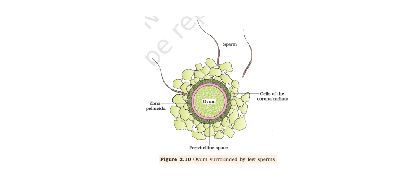
Sex Determination
- Female: The female is , so every ovum carries an chromosome.
- Male: The male is ; about half the sperms carry and half carry .
- Outcome: An -bearing sperm forms an zygote; a -bearing sperm forms an zygote.
- Chromosomal sex: The sperm determines whether the zygote is or ; therefore, the father determines the baby’s chromosomal sex.
| Sperm chromosome | Zygote | Development |
|---|---|---|
| Female | ||
| Male |
Cleavage and Implantation
Cleavage — A series of mitotic divisions of the zygote as it moves through the isthmus of the oviduct towards the uterus.
Early Embryo Stages
- Blastomeres: Cleavage produces daughter cells in the sequence 2, 4, 8 and 16; each cell is called a blastomere.
- Morula: The embryo with 8–16 blastomeres is called a morula.
- Blastocyst: Continued division forms a blastocyst with an outer trophoblast and an inner cell mass.
- Cell fates: The trophoblast attaches to the endometrium; the inner cell mass develops into the embryo.
| Stage | Key feature |
|---|---|
| Morula | Embryo with 8–16 blastomeres. |
| Blastocyst | Has an outer trophoblast and an inner cell mass. |
Implantation — The embedding of the blastocyst in the uterine endometrium.
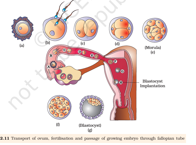
Placenta and Pregnancy Hormones
Placenta — A structural and functional unit formed by interdigitating chorionic villi and uterine tissue, linking the developing foetus with the mother.
| Hormone | Source or pregnancy-related point |
|---|---|
| hCG and hPL | Produced by the placenta; occur only during pregnancy. |
| Relaxin | Secreted by the ovary in the later phase; occurs only during pregnancy. |
| Estrogens and progestogens | Placenta produces these; maternal levels rise during pregnancy. |
| Cortisol, prolactin and thyroxine | Maternal blood levels increase severalfold during pregnancy. |
Germ Layers and Embryonic Development
- Germ layers: Soon after implantation, the inner cell mass forms outer ectoderm and inner endoderm; mesoderm appears between them.
- Importance: The three germ layers give rise to all tissues and organs of the adult body.
- Stem cells: The inner cell mass contains cells with the potency to form all tissues and organs.
- Pregnancy duration: Human pregnancy lasts about 9 months.
| Pregnancy stage | Major feature |
|---|---|
| After 1 month | The embryo’s heart has formed; its sound can be detected carefully with a stethoscope. |
| By the end of 2 months | Limbs and digits have developed. |
| By 12 weeks (end of first trimester) | Most major organ systems are formed; limbs and external genital organs are well developed. |
| Around the 5th month | Foetal movements are usually noticed and hair appears on the head. |
| By about 24 weeks (end of second trimester) | The body is covered with fine hair; eyelids separate and eyelashes form. |
| By the end of 9 months | The foetus is fully developed and ready for delivery. |
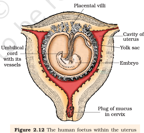
Do not confuse the morula with the blastocyst: the morula has 8–16 blastomeres, while the blastocyst has a trophoblast and an inner cell mass.
Where does fertilisation normally occur, and how many chromosomes are in the human zygote?
What are the main parts of a blastocyst, and what does each contribute to?
Quick recap
- Fertilisation occurs in the ampullary region when sperm and ovum arrive there simultaneously.
- Sperm entry blocks additional sperms and triggers completion of the secondary oocyte’s meiosis; fusion forms a 46-chromosome zygote.
- Cleavage begins as the zygote moves through the isthmus of the oviduct; divisions form blastomeres, then a morula and a blastocyst.
- Implantation embeds the blastocyst in the endometrium and leads to pregnancy.
- The placenta exchanges gases, nutrients and wastes, connects to the embryo by the umbilical cord, and produces pregnancy hormones.
- Ectoderm, mesoderm and endoderm give rise to body tissues and organs; foetal development proceeds through milestones until delivery at about nine months.
Parturition And Lactation
Gestation and Parturition
Oxytocin is injected to induce or strengthen uterine contractions during delivery.
Lactation
Explain the neuroendocrine mechanism of parturition.
What is colostrum, and why is it important?
Quick recap
- Human gestation lasts about nine months on average.
- Parturition is childbirth brought about by strong uterine contractions.
- Foetal and placental signals initiate the foetal ejection reflex.
- Oxytocin drives a positive-feedback cycle that strengthens uterine contractions.
- The placenta is expelled soon after the baby is delivered.
- Colostrum is antibody-rich first milk, and early breast-feeding is recommended.
Biology — ALL CHAPTERS
1
Sexual Reproduction In Flowering Plants
2
Human Reproduction
3
Reproductive Health
4
Principles Of Inheritance And Variation
5
Molecular Basis Of Inheritance
6
Evolution
7
Human Health And Disease
8
Microbes In Human Welfare
9
Biotechnology : Principles And Processes
10
Biotechnology And Its Applications
11
Organisms And Populations
12
Ecosystem
13
Biodiversity And Conservation