Class 12 PCB . Biology . Biology . Molecular Basis of Inheritance
Chapter 5: Molecular Basis Of Inheritance
Ch 5
Biology
Class 12 PCB
The DNA
Nucleic Acids and Genetic Material
DNA and RNA
- Nucleic acids are nucleotide polymers; DNA and RNA are the two types found in living systems.
- DNA is the genetic material in most organisms. RNA is genetic material in some viruses and mainly functions as a messenger.
- RNA roles also include adapter, structural and, in some cases, catalytic functions.
DNA Length
DNA length — The length of DNA is measured by the number of nucleotides in a single strand or base pairs in double-stranded DNA; it is characteristic of an organism.
| Organism | DNA length |
|---|---|
| Bacteriophage φX174 | 5,386 nucleotides |
| Bacteriophage lambda | 48,502 bp |
| Escherichia coli | 4.6 × 10⁶ bp |
| Human haploid DNA | 3.3 × 10⁹ bp |
Structure of a Polynucleotide Chain
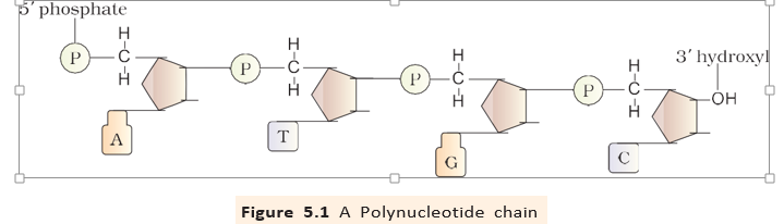
Nucleotide — A nucleotide consists of a nitrogenous base, a pentose sugar and a phosphate group.
| Class | Bases |
|---|---|
| Purines | Adenine (A), Guanine (G) |
| Pyrimidines | Cytosine (C), Thymine (T), Uracil (U) |
Building the Polynucleotide
- Sugar is deoxyribose in DNA and ribose in RNA. RNA ribose has an additional -OH group.
- Bases attach to the sugar's carbon by an N-glycosidic bond, forming a nucleoside.
- Nucleotide formation occurs when phosphate attaches to the sugar's -OH by a phosphoester bond.
- Chain formation links nucleotides through – phosphodiester bonds; the sugar and phosphate form the backbone.
- Ends are chemically different: the end has a free phosphate, while the end has a free OH group.
| Point | DNA | RNA |
|---|---|---|
| Sugar | Deoxyribose | Ribose, with a -OH group |
| Base difference | Contains thymine (T) | Uracil (U) replaces thymine |
DNA Double-Helix Model
Key Discoveries
- 1869Friedrich Meischer identified an acidic substance in the nucleus and named it “Nuclein”.
- 1953James Watson and Francis Crick proposed the DNA double-helix model using X-ray diffraction data from Maurice Wilkins and Rosalind Franklin.
Base Pairing and Complementarity
- Chargaff's observation: in double-stranded DNA, the amount of adenine equals thymine, and guanine equals cytosine.
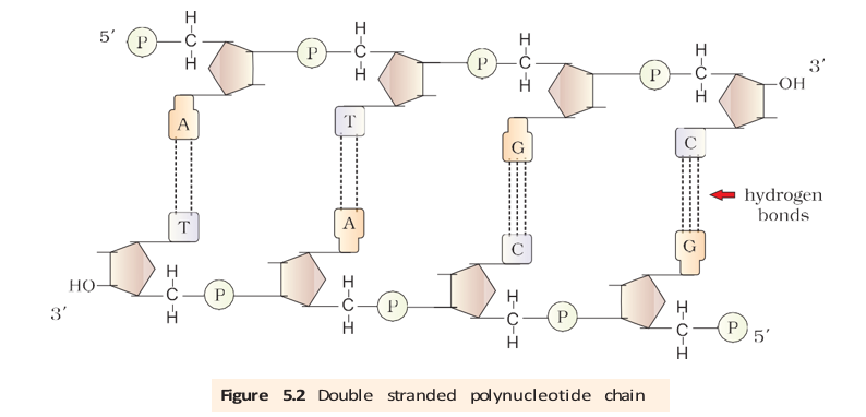
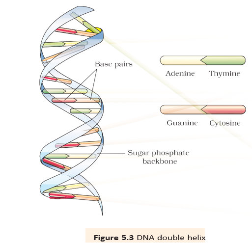
- Pairing: adenine pairs with thymine through two hydrogen bonds; guanine pairs with cytosine through three.
- Purine–pyrimidine pairing keeps the distance between the two strands approximately uniform.
- Complementary strands have predictable sequences: knowing one strand determines the sequence of the other.
- Template property: each parental strand can guide synthesis of a complementary strand, producing daughter DNA molecules identical in sequence to the parent.
For double-stranded DNA.
Salient Features of the Double Helix
- Two strands form the DNA molecule; sugar–phosphate backbones lie outside and the bases project inward.
- Antiparallel polarity means one strand runs to , while the other runs to .
- Complementary base pairs are held together by hydrogen bonds: A–T and G–C.
- Stability is supported by hydrogen bonding and the stacking of one base-pair plane over the next.
DNA and the Flow of Genetic Information
Central Dogma Preview
- DNA replicates to make DNA.
- DNA is transcribed to form mRNA.
- mRNA is translated to form protein.
State Chargaff's rules and explain how they relate to complementary base pairing.
What does antiparallel polarity mean in DNA?
Quick recap
- DNA is a nucleotide polymer and the genetic material of most organisms; RNA has genetic and several other roles.
- A nucleotide contains a base, pentose sugar and phosphate; nucleotides join by – phosphodiester bonds.
- DNA has deoxyribose and thymine, whereas RNA has ribose with a -OH group and uracil.
- DNA strands are antiparallel and complementary: A pairs with T, while G pairs with C.
- Hydrogen bonds and base stacking contribute to the stability of the double helix.
- Complementarity lets each DNA strand act as a template for producing an identical DNA molecule.
Packaging Of DNA Helix
How DNA Is Packaged
Multiply the number of base pairs by the distance between consecutive base pairs.
For example, mm of DNA corresponds to about base pairs.
Packaging in Prokaryotes and Eukaryotes
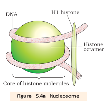
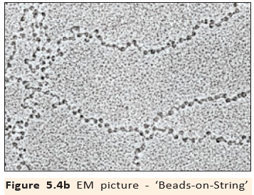
A mammalian cell with base pairs would contain about nucleosomes if each contains base pairs.
Chromatin and Higher-Level Packaging
| Feature | Euchromatin | Heterochromatin |
|---|---|---|
| Packing | Loosely packed | Densely packed |
| Staining | Light staining | Dark staining |
| Transcription | Transcriptionally active | Transcriptionally inactive |
The Search for Genetic Material
- Genetic material: Identifying the molecule responsible for inheritance became a major biological question as research advanced to the molecular level.
- Chromosomes: Earlier work had narrowed the search to chromosomes, but the molecule carrying hereditary information still needed to be established.
Griffith’s Transforming Principle
| Bacterial material injected | Effect on mice |
|---|---|
| Live S strain | Die |
| Live R strain | Live |
| Heat-killed S strain | Live |
| Heat-killed S strain + live R strain | Die; living S bacteria recovered |
Interpretation of Griffith’s Experiment
- Material from heat-killed S bacteria transferred to live R bacteria
- R bacteria acquired the ability to make a polysaccharide coat
- R bacteria became virulent S bacteria
Transforming principle — The substance from heat-killed S bacteria that changed live R bacteria into virulent S bacteria; Griffith did not identify its biochemical nature.
Biochemical Identification of the Transforming Principle
| Treatment | Effect on transformation | Inference |
|---|---|---|
| Protease digests protein | Transformation continued | Protein was not the transforming substance |
| RNase digests RNA | Transformation continued | RNA was not the transforming substance |
| DNase digests DNA | Transformation was inhibited | DNA was required for transformation |
DNase — An enzyme that digests DNA; unlike DNA, it is not the hereditary molecule itself.
What did Griffith’s experiment show?
How did Avery, MacLeod and McCarty show that DNA was the transforming principle?
Quick recap
- A mammalian cell’s DNA is about m long but must fit inside a tiny nucleus.
- Prokaryotic DNA forms protein-held loops in the nucleoid; eukaryotic DNA wraps around histone octamers to form nucleosomes.
- Nucleosomes form chromatin, which is further coiled and condensed into chromosomes.
- Euchromatin is loosely packed and active; heterochromatin is densely packed and inactive.
- Griffith showed that a transforming principle could convert live R bacteria into virulent S bacteria.
- Avery, MacLeod and McCarty found that DNA is required for transformation because DNase prevents it.
The Genetic Material Is DNA
Hershey–Chase Experiment
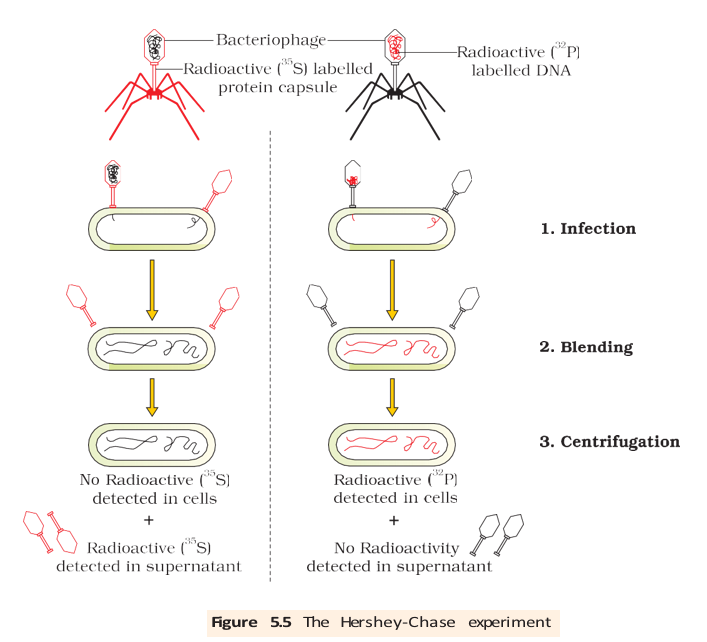
- Conclusion: DNA is the genetic material passed from bacteriophage to bacteria.
Properties of Genetic Material: DNA and RNA
Why DNA Is the Predominant Genetic Material
- Both can serve: DNA and RNA meet the requirements of replication, mutation and expression, but DNA is more stable.
- DNA stability: Its lower chemical reactivity and double-stranded complementary structure help preserve information; thymine also contributes to DNA stability.
- Repair: Complementary DNA strands help resist changes, and DNA has repair processes.
- RNA genomes: Some viruses use RNA as genetic material, including Tobacco Mosaic Virus and Qβ bacteriophage.
- Overall: DNA is preferred for long-term information storage, while RNA is better suited to information transmission.
Do not conclude that DNA is the only genetic material: RNA serves this role in some viruses, although DNA is the predominant and more stable genetic material.
RNA World
How did Hershey and Chase show that DNA, not protein, enters bacteria?
State the main properties required of genetic material.
Why is DNA generally preferred over RNA for storing genetic information?
Quick recap
- Hershey–Chase (1952): Radioactive phosphorus tracked DNA into bacteria; radioactive sulfur showed phage protein did not enter.
- Genetic material: Must replicate, remain stable, permit mutation and express inherited characters.
- DNA vs RNA: Both can be genetic material, but DNA is chemically and structurally more stable.
- RNA genomes: Tobacco Mosaic Virus and Qβ bacteriophage are examples of RNA-containing genetic material.
- RNA world: RNA likely came first, functioning as both genetic material and catalyst; DNA later evolved as a more stable information store.
Replication
Semiconservative DNA Replication
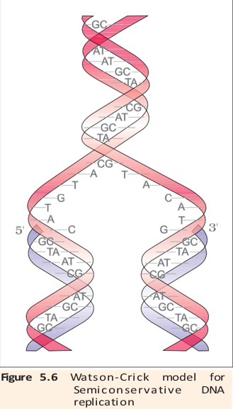
Experimental Proof: Meselson–Stahl Experiment
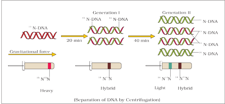
| Time / generation | DNA bands and proportions |
|---|---|
| After 20 minutes / 1 generation | All DNA is hybrid (intermediate density): one strand and one strand. |
| After 40 minutes / 2 generations | Equal amounts of hybrid and light DNA. |
| After 80 minutes / 4 generations | hybrid DNA and light DNA. |
Corroborating Evidence
- Further evidence: Taylor and colleagues used radioactive thymidine in faba bean (*Vicia faba*) chromosomes in 1958; their results also supported semiconservative replication.
- Conclusion: Semiconservative replication was demonstrated first in *E. coli* and later in higher organisms, including plants and human cells.
Enzymes, Energy and Replication Speed
- DNA polymerase: DNA-dependent DNA polymerase uses a DNA template to join deoxynucleotides; replication requires high speed and accuracy.
- Replication rate: *E. coli* copies its 4.6 × 10⁶ base pairs in about 18 minutes, at an average rate of roughly 2,000 base pairs per second.
- Energy: Deoxyribonucleoside triphosphates act as substrates and provide energy for polymerisation; other enzymes also help ensure accurate replication.
Replication Fork and Strand Synthesis
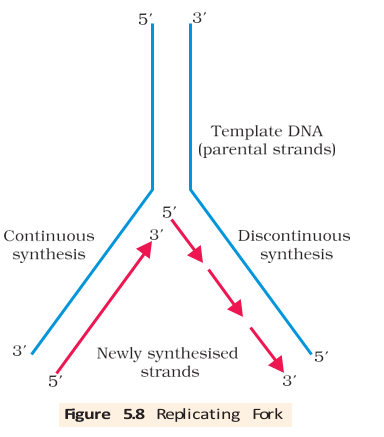
Origin of Replication and Cell-Cycle Timing
What does semiconservative replication mean?
What did Meselson and Stahl observe after one and two generations in medium?
Why is synthesis continuous on one template but discontinuous on the other?
Quick recap
- Semiconservative replication produces daughter DNA molecules with one old strand and one new strand.
- Meselson and Stahl used , and density-gradient centrifugation to demonstrate the process.
- After four generations in light nitrogen, the DNA is hybrid and light.
- DNA polymerase uses DNA templates, requires dNTPs and synthesises only in the direction.
- At the replication fork, synthesis is continuous on one template and discontinuous on the other; ligase joins the sections.
- Replication begins at an origin; eukaryotic DNA replication occurs in S phase and must be coordinated with cell division.
Transcription
Transcription and Its Direction
Transcription — The copying of genetic information from one DNA strand into an RNA molecule.
Why transcription copies only one DNA strand
- Complementary pairing: RNA is made by complementary base pairing; adenine pairs with uracil rather than thymine.
- One segment only: Unlike DNA replication, transcription copies only a particular DNA segment, not the entire DNA molecule.
- Different proteins: If both strands were transcribed, they could produce RNAs with different sequences and specify different proteins from the same DNA segment.
- Double-stranded RNA: The complementary RNAs could pair with each other, preventing their translation into proteins.
- RNA polymerase reads the template strand and synthesises RNA .
- RNA matches the coding strand except that uracil replaces thymine.
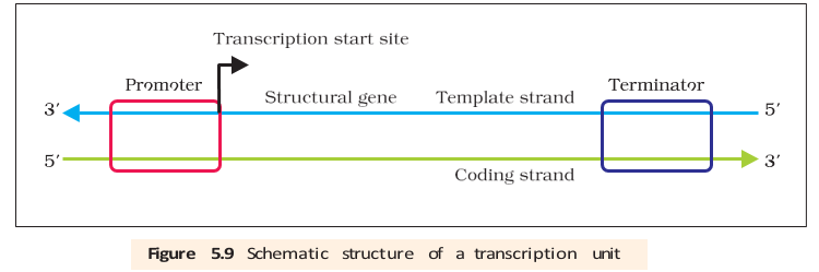
Transcription Unit
Transcription unit — A DNA region defined by a promoter, a structural gene and a terminator.
| Part | Position / role |
|---|---|
| Promoter | DNA sequence where RNA polymerase binds; lies upstream, toward the coding strand's end. |
| Structural gene | DNA region transcribed into RNA. |
| Terminator | Usually marks the end of transcription; lies downstream, toward the coding strand's end. |
| Point | Template strand | Coding strand |
|---|---|---|
| Direction | ||
| Role | Read by RNA polymerase to build complementary RNA. | Displaced during transcription; its sequence matches RNA except that DNA has thymine instead of uracil. |
| Naming | Also called the non-coding strand. | Called the coding strand, although it is not the strand used as the transcription template. |
Strand selection and RNA sequence
- Strand choice: Promoter position determines which DNA strand is the template; switching promoter and terminator positions reverses the strand assignments.
- Regulatory sequences: Additional sequences may occur upstream or downstream of the promoter and help regulate gene expression.
- Example: Template DNA produces RNA .
Transcription Unit and the Gene
Gene — A functional unit of inheritance; DNA sequences coding for tRNA or rRNA also count as genes.
| Term / type | Meaning |
|---|---|
| Cistron | A DNA segment that codes for a polypeptide. |
| Monocistronic | Structural gene with one cistron; typical of eukaryotes. |
| Polycistronic | Structural gene with multiple cistrons; common in bacteria and other prokaryotes. |
| Exons | Expressed sequences retained in mature or processed RNA. |
| Introns | Intervening sequences removed during RNA processing and absent from mature RNA. |
- Split genes: Eukaryotic monocistronic genes commonly contain coding exons interrupted by introns.
- Gene boundaries: Defining a gene only as a DNA segment is difficult because genes may encode RNA, and regulatory sequences also influence inheritance of a character.
- Regulatory genes: Promoter and other regulatory sequences are sometimes loosely called regulatory genes, even though they do not code for RNA or protein.
RNA Types and the Process of Transcription
| RNA | Role in protein synthesis |
|---|---|
| mRNA (messenger RNA) | Provides the template carrying the genetic message. |
| tRNA (transfer RNA) | Brings amino acids and reads the genetic code. |
| rRNA (ribosomal RNA) | Has structural and catalytic roles in translation. |
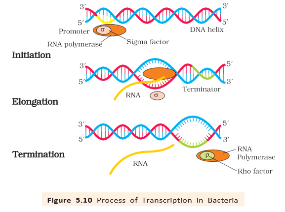
Bacterial transcription
- One polymerase: A single DNA-dependent RNA polymerase transcribes all three major RNA types.
- Substrates and synthesis: RNA polymerase uses nucleoside triphosphates, follows template complementarity, helps open the DNA helix and synthesises RNA in the direction.
- Initiation: The enzyme associates transiently with initiation factor ; this changes its specificity and enables initiation at the promoter.
- Elongation: RNA polymerase itself catalyses elongation; only a short stretch of the new RNA remains bound to the enzyme.
- Termination: Transient association with termination factor alters enzyme specificity and enables termination; nascent RNA and polymerase are released at the terminator.
- Coupling: Bacterial mRNA needs no processing, and transcription and translation occur in the same compartment. Translation can begin before transcription is complete.
Eukaryotic RNA polymerases
- Polymerase count: Eukaryotic nuclei have at least three RNA polymerases; additional RNA polymerases are present in organelles.
- Division of labour: The three nuclear polymerases transcribe different RNA types.
| Polymerase | Main RNA products |
|---|---|
| RNA polymerase I | rRNAs , and . |
| RNA polymerase II | Precursor of mRNA, called heterogeneous nuclear RNA (hnRNA). |
| RNA polymerase III | tRNA, rRNA and small nuclear RNAs (snRNAs). |
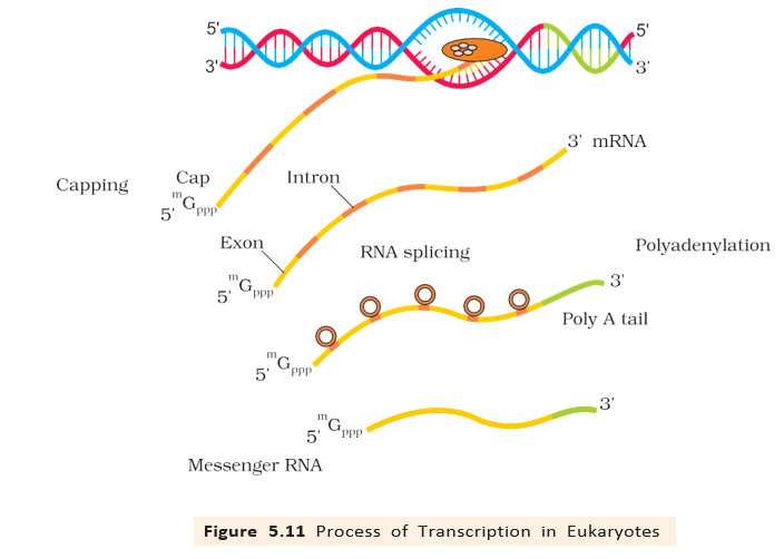
Eukaryotic hnRNA processing
- Primary transcript: hnRNA contains both exons and introns and is not yet functional mRNA.
- Splicing: Introns are removed and exons are joined in the correct order.
- Capping: An unusual methyl guanosine triphosphate nucleotide is added to the end.
- Tailing: About 200–300 adenylate residues are added to the end without using a template.
- Mature mRNA: After processing, hnRNA becomes mRNA and is transported out of the nucleus for translation.
Why is only one DNA strand transcribed?
Distinguish the coding and template strands.
What processing converts eukaryotic hnRNA into mRNA?
Quick recap
- Transcription copies one DNA segment from one template strand into RNA.
- The transcription unit contains a promoter, structural gene and terminator.
- RNA matches the coding strand except that RNA uses uracil instead of thymine.
- A cistron codes for a polypeptide; eukaryotic genes often contain exons separated by introns.
- Bacteria use one RNA polymerase: it catalyses elongation, while transient and associations support initiation and termination.
- Eukaryotic nuclei have at least three RNA polymerases, with additional polymerases in organelles; hnRNA is processed into mature mRNA.
Genetic Code
Meaning and deciphering of the genetic code
Genetic code — The genetic code is the set of rules by which mRNA nucleotide sequences specify the amino-acid sequence of a protein.
Why a code is needed
- Translation: Transfers genetic information from a nucleotide polymer to an amino-acid polymer; no direct complementarity links nucleotides to amino acids.
- George Gamow: Proposed that combinations of bases encode amino acids. Four bases in triplets give possible codons—enough for 20 amino acids.
- Har Gobind Khorana: Developed methods to synthesise RNA molecules with defined base combinations, including homopolymers and copolymers.
- Marshall Nirenberg: His cell-free protein-synthesis system helped decipher the code.
- Severo Ochoa: Polynucleotide phosphorylase helped synthesise RNA without a template.
Codon chart
How to read the chart
- Direction: Read each mRNA codon from to .
- Selection: Use the first base to select a row group, the second base to select a column, and the third base to identify the codon within that group.
| First two bases | Third base | Third base | Third base | Third base |
|---|---|---|---|---|
| — phenylalanine | — phenylalanine | — leucine | — leucine | |
| — serine | — serine | — serine | — serine | |
| — tyrosine | — tyrosine | — stop | — stop | |
| — cysteine | — cysteine | — stop | — tryptophan | |
| — leucine | — leucine | — leucine | — leucine | |
| — proline | — proline | — proline | — proline | |
| — histidine | — histidine | — glutamine | — glutamine | |
| — arginine | — arginine | — arginine | — arginine | |
| — isoleucine | — isoleucine | — isoleucine | — methionine; start | |
| — threonine | — threonine | — threonine | — threonine | |
| — asparagine | — asparagine | — lysine | — lysine | |
| — serine | — serine | — arginine | — arginine | |
| — valine | — valine | — valine | — valine | |
| — alanine | — alanine | — alanine | — alanine | |
| — aspartic acid | — aspartic acid | — glutamic acid | — glutamic acid | |
| — glycine | — glycine | — glycine | — glycine |
Properties of the genetic code
- Triplet: Each codon consists of three mRNA nucleotides. Of the 64 codons, 61 specify amino acids and three are stop codons.
- Degenerate: Several different codons can specify the same amino acid.
- Commaless: Codons are read continuously, without gaps or punctuation.
- Nearly universal: For example, specifies phenylalanine from bacteria to humans. Exceptions occur in mitochondrial codons and some protozoans.
- AUG: Specifies methionine and also acts as the initiator codon.
- Stop codons: , and terminate translation; they do not specify amino acids.
Reading and reversing a codon sequence
- Example: translates to methionine followed by six phenylalanines.
- Reverse prediction: Methionine followed by six phenylalanines cannot determine one unique mRNA sequence because phenylalanine has more than one codon; this demonstrates degeneracy.
Mutations and the genetic code
Point mutations and frameshifts
- Point mutation: A change in one base pair can alter the amino acid specified by a gene.
- Sickle-cell anaemia: A single base-pair change in the beta-globin gene changes glutamate to valine in the protein.
- Frameshift: Inserting or deleting one or two bases changes how the sequence is grouped into triplets from the mutation onward.
- Multiple of three: Inserting or deleting three bases, or a multiple of three, adds or removes one or more amino acids but leaves the downstream reading frame unchanged.
| Mutation | Effect on reading frame |
|---|---|
| One or two bases inserted/deleted | Reading frame shifts from the mutation site. |
| Three bases or a multiple of three inserted/deleted | One or more amino acids are added/removed; downstream frame remains unchanged. |
tRNA as the adapter molecule
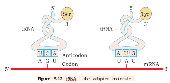
Adapter molecule — tRNA connects an mRNA codon to its corresponding amino acid during translation.
tRNA structure and function
- Anticodon loop: Carries bases complementary to the mRNA codon.
- Amino-acid acceptor end: Binds the specific amino acid carried by that tRNA; tRNAs are specific to each amino acid.
- Initiator tRNA: A distinct tRNA is used to begin translation.
- Stop codons: No tRNAs correspond to stop codons.
- Shape: The secondary structure is cloverleaf-like, while the folded three-dimensional molecule is compact and resembles an inverted L.
- Earlier name: tRNA was previously called soluble RNA (sRNA).
Why is the genetic code called degenerate?
What is the effect of inserting one base into a structural gene?
Quick recap
- Codons are triplets read continuously on mRNA; 61 specify amino acids and three signal termination.
- The code is degenerate and nearly universal, with exceptions in mitochondrial codons and some protozoans.
- AUG codes for methionine and serves as the initiator; UAA, UAG and UGA are stop codons.
- A beta-globin point mutation changing glutamate to valine causes sickle-cell anaemia.
- Insertions or deletions not in multiples of three shift the reading frame; multiples of three preserve it downstream.
- tRNA links codons and specific amino acids through its anticodon and amino-acid acceptor end; no tRNAs read stop codons.
Translation
Translation and Protein Synthesis
Translational unit — The mRNA sequence between a start codon and a stop codon that codes for a polypeptide.
Main Stages of Translation
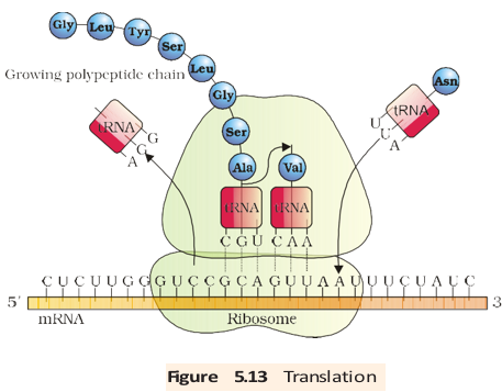
- Initiation: The ribosome binds mRNA at AUG, recognised by the initiator tRNA.
- Elongation: Charged tRNAs pair their anticodons with complementary mRNA codons. The ribosome moves along the mRNA and adds amino acids one at a time.
- Termination: A release factor binds to a stop codon, ending translation and releasing the completed polypeptide.
Regulation of Gene Expression
Gene expression regulation — Control of when and how strongly a gene is expressed to produce its functional product.
| Control feature | Key point |
|---|---|
| Main control point | Control of transcription initiation is the predominant point of gene regulation. |
| Accessory proteins | Activators and repressors affect RNA polymerase's ability to recognise transcription start sites. |
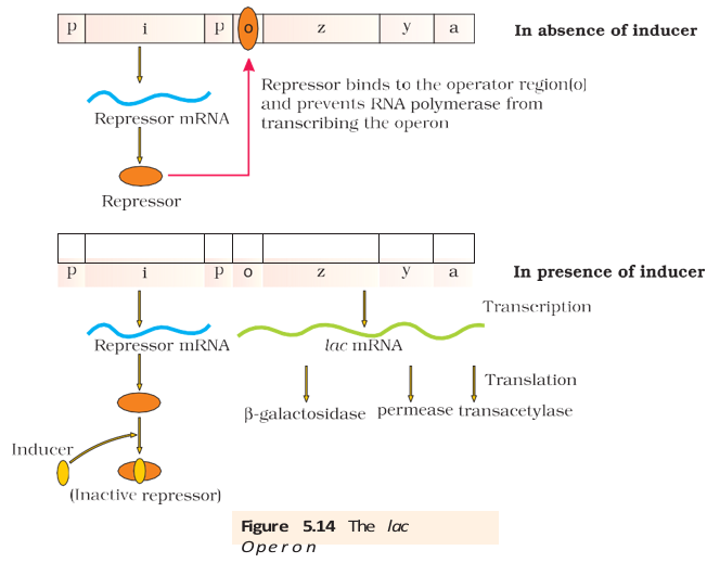
| Operator | A DNA sequence, usually beside the promoter, that regulates promoter accessibility through protein binding. |
| Specificity | Each operon has its own operator and specific repressor; the lac repressor binds the lac operator. |
The Lac Operon
Operon — A bacterial gene arrangement in which related structural genes are controlled together by a common promoter and regulatory elements.
| Condition | Without inducer | With inducer |
|---|---|---|
| Repressor | Binds the operator. | Lactose or allolactose binds to and inactivates the repressor. |
| RNA polymerase | Blocked from transcribing the operon. | Can access the promoter; transcription proceeds. |
| Operon state | Off: structural genes are not transcribed. | On: structural genes are transcribed. |
Induction and Regulation
- Inducer: Lactose, or allolactose, can inactivate the lac repressor and switch on the operon. Glucose and galactose are not inducers.
- Lactose entry: A low basal level of lac operon expression allows some permease to be made, enabling lactose to enter the cell.
- Negative regulation: The repressor controls the operon by preventing transcription when it occupies the operator.
- Positive regulation: The lac operon is also subject to positive regulation, but its details are beyond this lecture.
What is tRNA charging, and why is it necessary?
How does an inducer switch on the lac operon?
Quick recap
- Translation uses mRNA codons to specify the amino acid sequence of a polypeptide.
- ATP-dependent aminoacylation attaches amino acids to their cognate tRNAs.
- Translation proceeds through initiation, elongation and release-factor-mediated termination.
- Gene expression in eukaryotes can be regulated at transcription, RNA processing, transport and translation.
- In prokaryotes, transcription initiation is a major control point, influenced by activators and repressors.
- In the lac operon, lactose or allolactose inactivates the repressor, allowing transcription of genes needed for lactose metabolism.
Human Genome Project
Purpose and Scale of the Human Genome Project (HGP)
Why it was a mega project
- Scale: The human genome was estimated to contain about 3 billion base pairs; at the initial estimate of 3 US dollars per base pair, sequencing would cost about 9 billion US dollars.
- Data storage: Printing the sequence at 1,000 letters per page and 1,000 pages per book would require about 3,300 books for one human cell.
- Computing: The volume of data required high-speed computer systems for storage, retrieval and analysis.
- Bioinformatics: HGP was closely linked to the development of this field, which applies computational tools to biological data.
Goals, Coordination and Outcomes
Major goals
- Identify genes: Find all human genes; the initial goal estimated about 20,000–25,000 genes.
- Sequence DNA: Determine the sequence of the 3 billion chemical base pairs in human DNA.
- Manage data: Store sequence information in databases and improve tools for analysing it.
- Share technology: Transfer related technologies to other sectors, including industry.
- Address ELSI: Consider the ethical, legal and social issues arising from the project.
Coordination and significance
- Leadership: A 13-year project coordinated by the U.S. Department of Energy and the National Institutes of Health.
- Partners: Wellcome Trust (U.K.) became a major partner; Japan, France, Germany, China and other countries also contributed.
- Completion: The project was completed in 2003.
- Importance: Understanding DNA variation can improve diagnosis and treatment, and may help prevent many disorders.
- Wider value: Sequencing non-human organisms can reveal useful capabilities for healthcare, agriculture, energy production and environmental remediation.
- Other genomes: Sequenced organisms included bacteria, yeast, *Caenorhabditis elegans*, fruit flies, rice and *Arabidopsis*.
Methodologies Used in HGP
| Approach | What it does | Key term |
|---|---|---|
| Gene-focused | Identifies genes that are expressed as RNA. | Expressed Sequence Tags (ESTs) |
| Whole-genome | Sequences coding and non-coding DNA without first selecting genes; functions are assigned later. | Sequence annotation |
Whole-genome sequencing and assembly
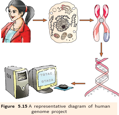
- Isolate total DNA from a cell.
- Break it into random, smaller fragments because very long DNA molecules are difficult to sequence directly.
- Clone fragments in bacteria or yeast using BACs (bacterial artificial chromosomes) or YACs (yeast artificial chromosomes); cloning amplifies each fragment.
- Sequence the fragments with automated sequencers based on Frederick Sanger’s method.
- Use overlaps between fragments to assemble their sequences with specialised computer programs.
- Annotate the assembled sequence, assign regions to chromosomes, and construct genetic and physical maps using restriction-site polymorphisms and microsatellites.
Mapping and sequencing details
- Sequence assembly: Overlapping fragments allow separately sequenced pieces to be aligned; this task required computer programs.
- Genome maps: Restriction endonuclease recognition-site polymorphisms and repetitive DNA sequences called microsatellites helped generate genetic and physical maps.
- Chromosome 1: Its sequence was completed in May 2006, the last of the 24 human chromosomes to be sequenced (22 autosomes plus X and Y).
Salient Features of the Human Genome
- Genome size: The human genome contains 3,164.7 million base pairs.
- Gene size: An average gene has about 3,000 bases, but sizes vary greatly; the dystrophin gene is the largest known, at 2.4 million bases.
- Gene number: The project’s observations estimated about 30,000 genes, lower than earlier estimates of 80,000–140,000.
- Shared sequence: About 99.9 percent of nucleotide bases are identical among people.
- Unknown function: The functions of over 50 percent of discovered genes were unknown.
- Protein-coding DNA: Less than 2 percent of the genome codes for proteins.
- Repeated DNA: Repetitive sequences form a large portion of the genome. Though thought to have no direct coding function, they provide clues about chromosome structure, dynamics and evolution.
- Gene distribution: Chromosome 1 has the most genes (2,968); the Y chromosome has the fewest (231).
- SNPs: About 1.4 million locations of single-base differences were identified; they can help locate disease-associated sequences and trace human history.
Applications and Future Challenges
| Point | Earlier approach | Genome-wide approach |
|---|---|---|
| Scope | Researchers studied one or a few genes at a time. | Whole-genome sequences and new high-throughput technologies enable systematic study of all genes and their products on a much broader scale. |
| Examples | Focused analysis of selected genes. | Study all transcripts in a tissue, organ or tumour, or examine how many genes and proteins work together in interconnected networks. |
- Bioinformatics
- Use of computational methods to store, retrieve and analyse biological data.
- EST
- A tag used to identify genes that are expressed as RNA.
- Sequence annotation
- Assigning functions to different regions of a genome sequence.
- SNP
- Single nucleotide polymorphism: a variation at a single DNA base among individuals.
- Microsatellite
- A repetitive DNA sequence used in genome mapping.
What were the two major approaches used in HGP?
State four important findings of the Human Genome Project.
Quick recap
- HGP began in 1990 and was completed in 2003; it sequenced and analysed the human genome.
- Its goals included identifying genes, sequencing DNA, storing data, improving analysis, sharing technology and addressing ELSI.
- HGP used both expressed-gene identification (ESTs) and whole-genome sequencing followed by annotation.
- Small DNA fragments were cloned, sequenced, assembled using overlaps, computer-analysed and mapped to chromosomes.
- The genome has 3,164.7 million base pairs, but less than 2 percent is protein-coding DNA.
- Whole-genome sequences and high-throughput technologies support systematic study of genes, transcripts and gene-protein networks.
DNA Fingerprinting
DNA Variation and DNA Fingerprinting
DNA fingerprinting — A technique for identifying differences in DNA patterns among individuals, based mainly on highly variable repetitive DNA sequences.
Why DNA fingerprinting works
- Human DNA variation: About 99.9% of human DNA sequences are the same. For a genome of base pairs, the remaining 0.1% represents about differing base pairs.
- Repetitive DNA: Some regions contain short DNA sequences repeated many times; these regions vary greatly between individuals.
- Satellite DNA: During density-gradient centrifugation, repetitive DNA forms smaller satellite peaks, separate from the main bulk-DNA peak.
- Satellite classes: Microsatellites and minisatellites are classified by base composition, segment length and number of repeat units.
- Coding: Repetitive DNA usually does not code for proteins but forms a substantial part of the human genome.
DNA Polymorphism
DNA polymorphism — Heritable variation in DNA sequence; traditionally, a locus is considered polymorphic when more than one allele occurs in the human population at a frequency greater than 0.01.
How polymorphism arises
- Origin: Mutations produce DNA sequence variations, from single-nucleotide changes to large-scale changes.
- Somatic mutations: These occur in body cells and are generally not passed to offspring.
- Germ-cell mutations: These occur in cells that produce gametes; they can be inherited and spread through a population if they do not prevent reproduction.
- Non-coding DNA: Mutations here are more likely to persist because they may not immediately affect reproductive ability.
- Accumulation: Inherited variations can build up over generations, contributing to genetic variability and forming a basis for genetic mapping and DNA fingerprinting.
VNTRs and the DNA Fingerprinting Technique
Variable Number of Tandem Repeats (VNTRs)
- Development: Alec Jeffreys initially developed DNA fingerprinting using highly polymorphic satellite DNA as a probe.
- VNTR structure: A short DNA sequence is repeated in tandem many times; the repeat number varies between chromosomes and individuals.
- Classification: VNTRs are a type of minisatellite DNA.
- Fragment size: VNTR-containing fragments vary in size from 0.1 to 20 kb.
- Pattern: Different repeat numbers produce fragments of different sizes and, after detection, a characteristic band pattern for an individual.
Traditional Southern Blot DNA Fingerprinting
- Isolate DNA from the sample.
- Cut the DNA into fragments using restriction endonucleases.
- Separate the fragments by electrophoresis.
- Transfer the separated fragments onto a nitrocellulose or nylon membrane by blotting.
- Hybridise the fragments with a radiolabelled VNTR probe.
- Detect the hybridised fragments by autoradiography; the resulting bands form the DNA fingerprint.
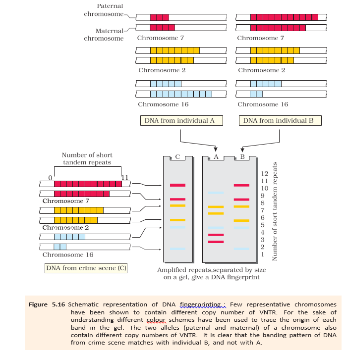
Reading and Uses of DNA Fingerprints
- Individual pattern: VNTR band patterns generally differ between people; monozygotic (identical) twins are an exception.
- Same person, different tissues: DNA from blood, hair follicles, skin, bone, saliva or sperm of one person shows the same degree of polymorphism.
- Forensics: Band-pattern matching can help identify individuals from biological samples.
- Paternity testing: Because polymorphisms are inherited, a child's DNA pattern can be compared with those of the parents.
- Other applications: DNA fingerprinting helps study population and genetic diversity, with applications in evolutionary biology.
- Improved sensitivity: PCR increased sensitivity so that DNA from a single cell can be sufficient; many different probes are now used.
Do not assume every person's DNA fingerprint is unique: monozygotic (identical) twins generally have the same pattern.
What is DNA polymorphism?
List the main steps in traditional DNA fingerprinting.
Quick recap
- DNA fingerprinting compares individuals using variable repetitive DNA regions, especially VNTRs.
- DNA polymorphism is heritable DNA variation; the traditional threshold is an allele frequency greater than 0.01.
- VNTR repeat-number differences create fragments of different lengths and characteristic band patterns.
- Traditional fingerprinting uses restriction digestion, electrophoresis, Southern blotting, radiolabelled-probe hybridisation and autoradiography.
- DNA fingerprinting is used in forensics, paternity testing and studies of genetic diversity.
- PCR makes the method sensitive enough to analyse DNA from a single cell.
Biology — ALL CHAPTERS
1
Sexual Reproduction In Flowering Plants
2
Human Reproduction
3
Reproductive Health
4
Principles Of Inheritance And Variation
5
Molecular Basis Of Inheritance
6
Evolution
7
Human Health And Disease
8
Microbes In Human Welfare
9
Biotechnology : Principles And Processes
10
Biotechnology And Its Applications
11
Organisms And Populations
12
Ecosystem
13
Biodiversity And Conservation