Class 9 . Science . Exploration . Cell the Building Block of Life
Chapter 2: Cell: The Building Block of Life
Ch 2
Science
Class 9
How to Study Cells?
Origin and Organisation of Cells
Possible Early Environments
- Early conditions: Life is widely thought to have originated in water; small pools with changing conditions, such as hot springs, may have supported early life.
- Early Earth: Hot-spring conditions may resemble those on Earth about billion years ago.
- Puga Valley: Hot springs in Ladakh remain near boiling even in a cold climate and contain unicellular, heat-loving bacteria called thermophiles.
- Possible protection: Researchers at the Birbal Sahni Institute of Palaeosciences, Lucknow, found rapid calcium carbonate deposits around the springs. These may have protected early organic molecules from radiation and extreme conditions.
- First membrane: The deposits may also have helped form the first protective membrane—the barrier that defines a cell.
Cell — The cell is the basic structural and functional unit of every living organism.
| Type | Description | Examples |
|---|---|---|
| Unicellular | Made of one cell. | Bacteria, yeast |
| Multicellular | Made of many cells that work together. | Plants, fish, birds, humans |
Organisation in Multicellular Organisms
- Similar cells performing similar functions form a tissue.
- Different tissues are organised into an organ.
- Several organs work together as an organ system; for example, nasal pores, the nasal cavity, trachea and lungs form the respiratory system.
Why the Cell Is Fundamental
- Structure: Cells make up tissues, organs and organ systems.
- Function: Cells perform the activities that keep organisms alive, individually or as part of a larger organism.
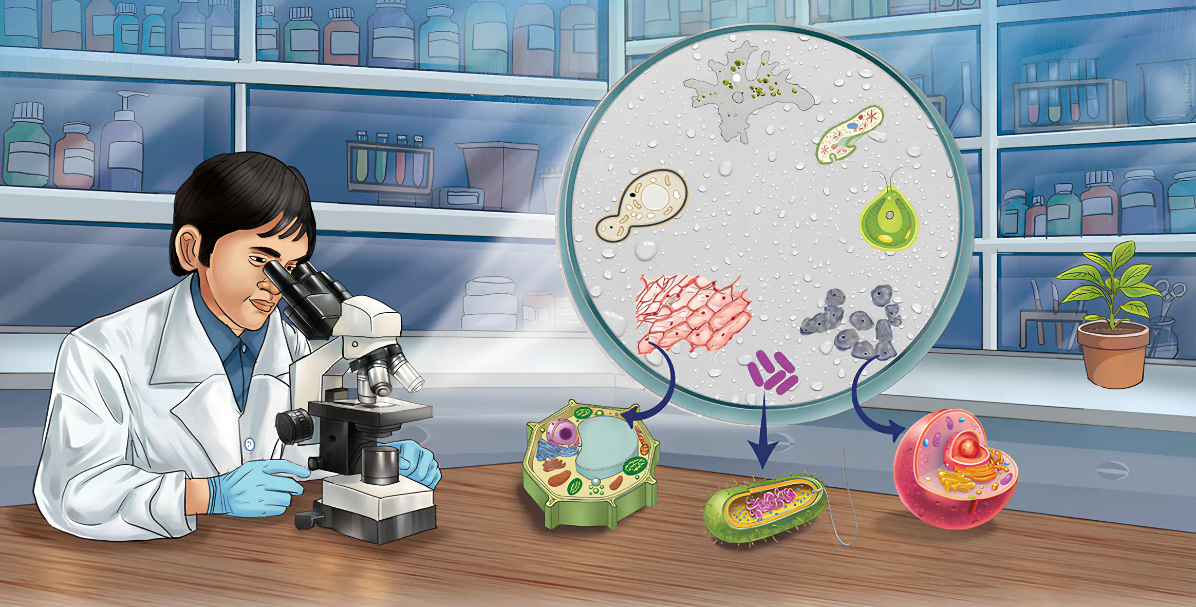
Seeing and Measuring Cells
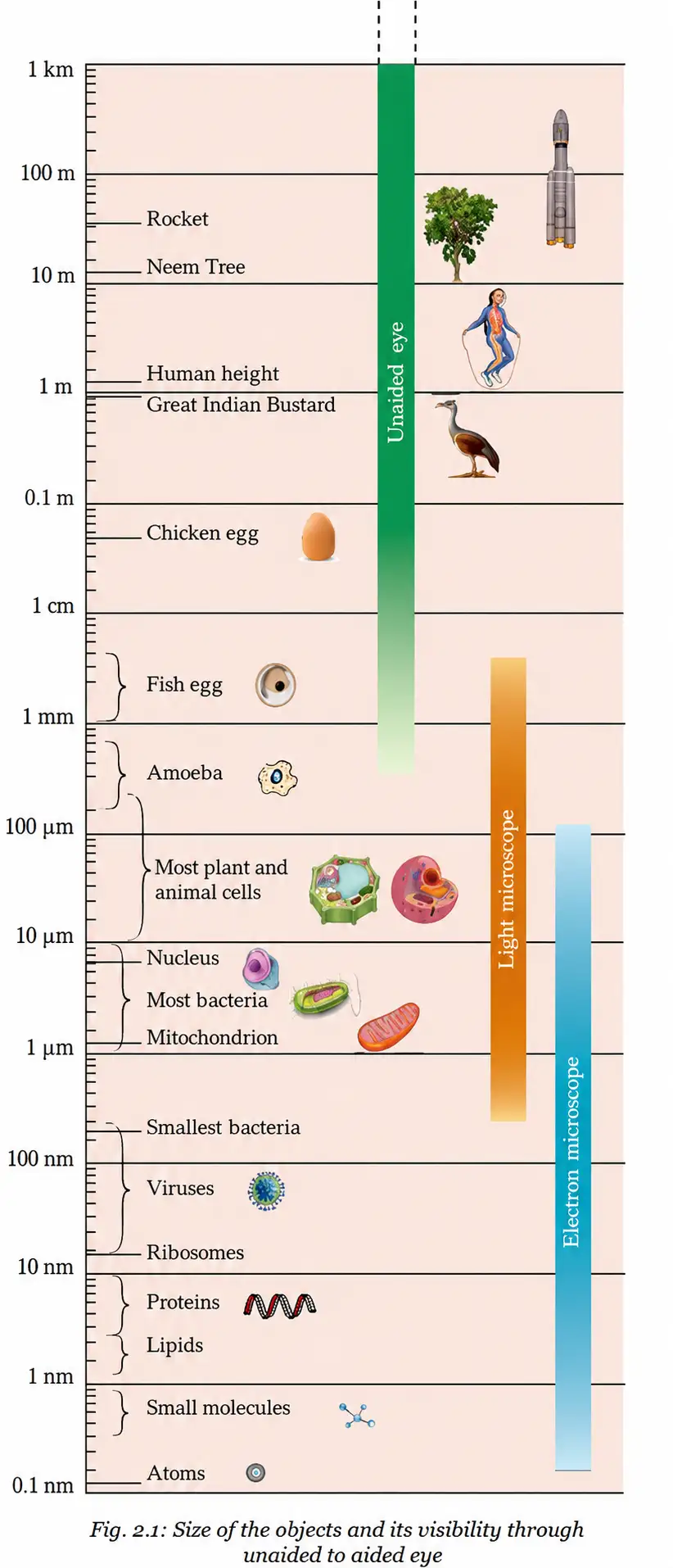
Limit of resolution — The limit of resolution is the smallest separation at which two nearby points can still be seen as distinct.
Human Vision and Cells
- Human eye: At its near point of about , the eye can distinguish points about apart.
- Cells: A cell is usually too small to be seen by the unaided eye, so microscopes are needed to study its structure.
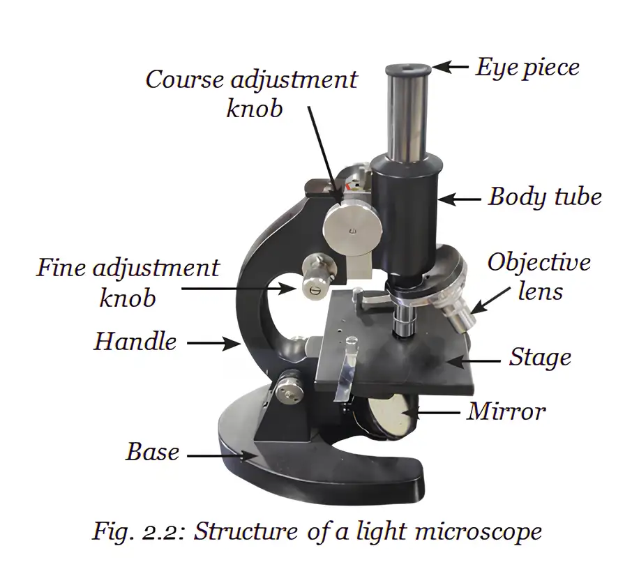
Microscopes and Magnification
- Lens action: A convex lens, or an objective lens and eyepiece together, magnifies an object so it appears larger.
- Robert Hooke: In 1665, he used a self-designed microscope with about – magnification to examine cork. He saw box-like compartments and named them “cells.”
- Light microscope: Uses visible light and lenses; school microscopes commonly have objective lenses such as and .
Convert the field-of-view diameter to micrometres, then divide it by the number of cells spanning the diameter. Since , convert millimetres before calculating.
Cell-Size Estimate Example
- Example: A field diameter of equals . If cells span it, one cell is about .
- Symbols: is the field-of-view diameter in micrometres; is the number of cells across it.
| Point | Light microscope | Electron microscope |
|---|---|---|
| Source used | Visible light | A beam of electrons |
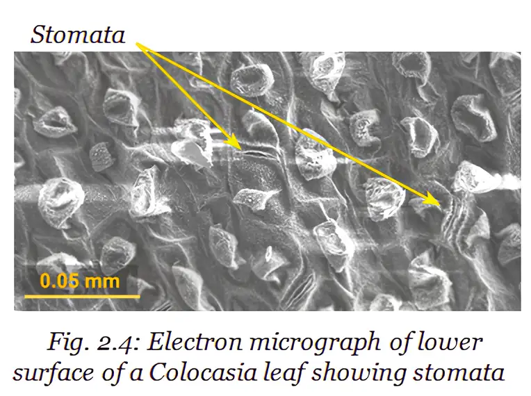
| Detail revealed | Magnified images of objects | Fine cell details, including structures at the nanometre scale |
| Example | Commonly used in school laboratories | Scanning electron microscope image of stomata on a Colocasia leaf |
Nanometre — A nanometre is one-billionth of a metre: .
Multiply the magnifying power of the eyepiece by that of the objective lens. For example, and give a total magnification of .
What Makes Microscopes Better?
- Magnification: How much larger the image appears.
- Resolution: The clarity with which fine details can be distinguished.
- Contrast: The difference in brightness between parts of an object.
What is the limit of resolution of the human eye?
How is cell size estimated from a microscope's field of view?
Quick recap
- Cells are the basic structural and functional units of living organisms.
- Organisms may be unicellular or multicellular; multicellular organisation proceeds from cells to tissues, organs and organ systems.
- Calcium carbonate deposits around hot springs may have protected early organic molecules and helped form the protective membrane that defines a cell.
- The human eye resolves points about apart under the stated viewing conditions; Hooke named cells after examining cork in 1665.
- Estimate cell size by dividing the field-of-view diameter in micrometres by the number of cells across it.
- Total magnification is the eyepiece magnification multiplied by the objective magnification.
- Electron microscopes use electrons to reveal finer cell details; microscopes improve through magnification, resolution and contrast.
Structure of a Cell
Cell Membrane and Movement of Substances
Diffusion — The net movement of particles from a region of higher concentration to a region of lower concentration, with or without a membrane.
Osmosis — The diffusion of water through a selectively permeable membrane, from a dilute solution (more water) towards a concentrated solution (less water).
Osmosis in cells
- Direction: Water moves across the membrane until the concentrations on the two sides become equal.
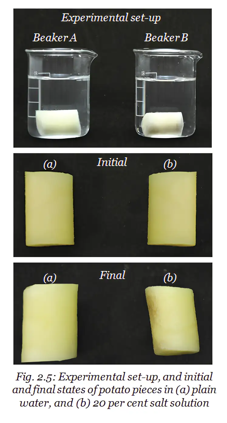
- Potato in plain water: Water enters the cells by osmosis, so the piece swells and gains weight.
- Potato in concentrated salt or sugar solution: Water leaves the cells, so the piece shrinks and loses weight.
- In plants: Water enters root cells from the soil by osmosis.
- Prediction: Soaked mung bean seeds placed in a concentrated solution lose water and shrink.
| Solution outside cell | Relative concentration | Water movement | Effect on cell |
|---|---|---|---|
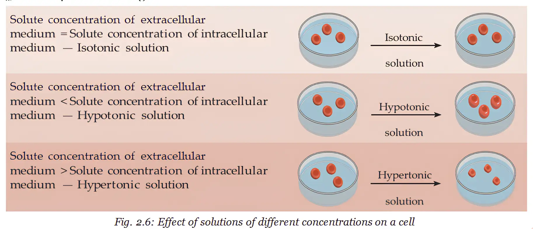
| Hypotonic | More dilute than the cell | Water enters | Cell swells |
| Isotonic | Equal to the cell | No net water movement | Cell size remains about the same |
| Hypertonic | More concentrated than the cell | Water leaves | Cell shrinks |
Cell Membrane Structure
Cell Wall
| Point | Plant cells | Animal cells |
|---|---|---|
| Cell wall | Present outside the cell membrane | Absent |
| Shape and support | Rigid wall helps maintain shape and firmness | Without a rigid wall, cells can change shape more easily |
| In concentrated sugar solution | Water leaves; inner contents shrink away from the wall, but the wall maintains the cell’s outer shape | Water leaves and the cell shrinks |
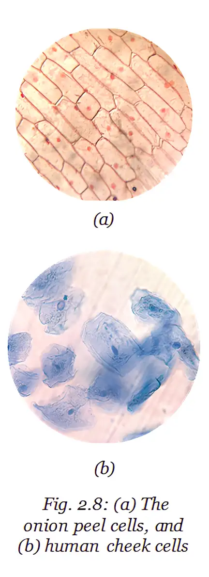
| Example in the lecture | Onion peel or Rhoeo leaf cells are box-shaped and regularly arranged | Cheek cells are irregularly arranged |
Osmosis and cell walls
- Plant cells in concentrated sugar solution: Water leaves by osmosis; the cell membrane and inner contents pull away from the rigid wall.
- Plant cell shape: The cell’s contents shrink, but the outer wall helps maintain the cell’s shape.
- Animal cells: Without a cell wall, they shrink when they lose water and can change shape more easily.
- Cell flexibility: The flexible shape of animal cells supports movement and the functioning of animal tissues.
- Other organisms: Fungi and bacteria also have cell walls for protection and structural support.
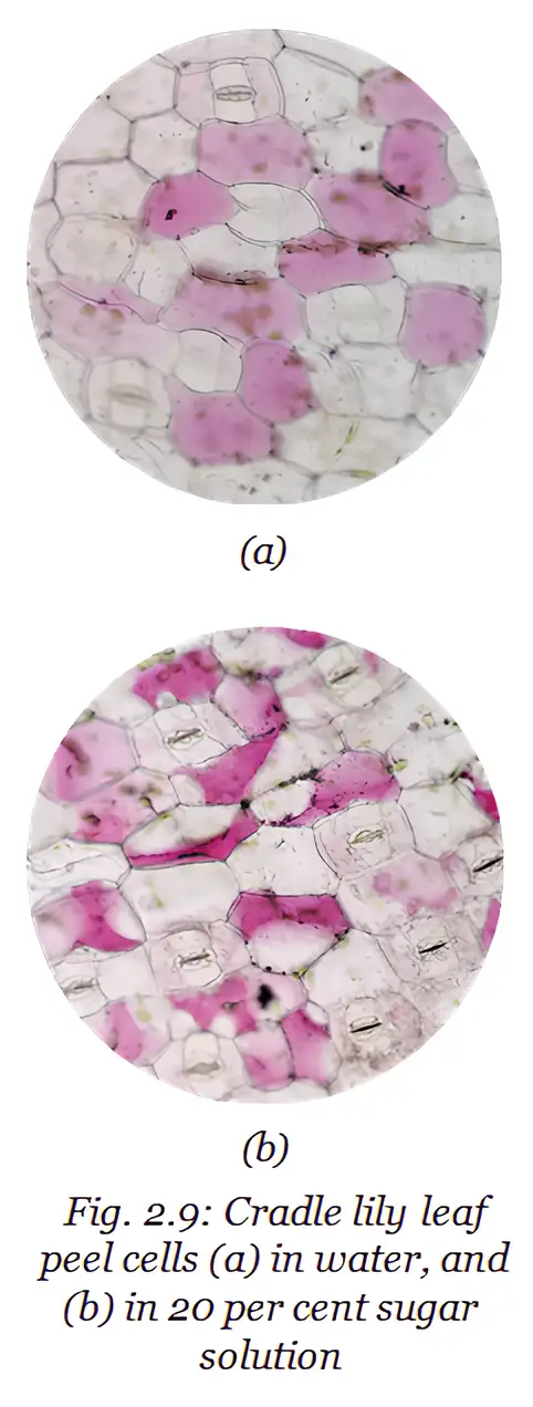
A plant cell’s contents may shrink in a concentrated solution, but its rigid cell wall helps preserve the cell’s outer shape; animal cells lack this wall and shrink more directly.
Why do plant cells need a cell wall?
How do plant and animal cells differ in a concentrated sugar solution?
Quick recap
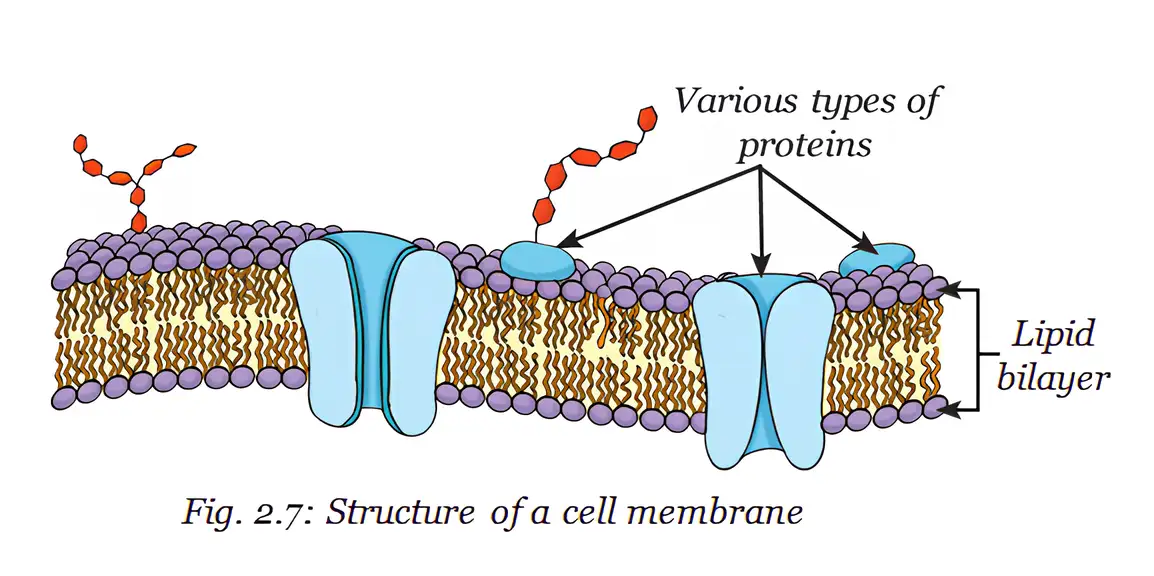
- The selectively permeable cell membrane controls exchange between a cell and its surroundings.
- Diffusion moves particles from higher to lower concentration; osmosis is water diffusion through a selectively permeable membrane.
- Water enters cells in hypotonic solutions and leaves them in hypertonic solutions; isotonic solutions cause no net water movement.
- The fluid-mosaic model describes a moving lipid bilayer with embedded proteins.
- Plant, fungal and bacterial cells have a cell wall; plant walls are mainly cellulose.
- Plant cell walls provide support and maintain shape, while animal cells lack a wall and can change shape more easily.
The Cell Interior - A Coordinated Working System
Basic cell organisation
Main parts
- Plasma membrane: A selectively permeable boundary around the cell.
- Cytoplasm: A semi-fluid, jelly-like substance containing organelles and other materials.
- Organelles: Specialised structures in the cytoplasm; many are visible only with an electron microscope.
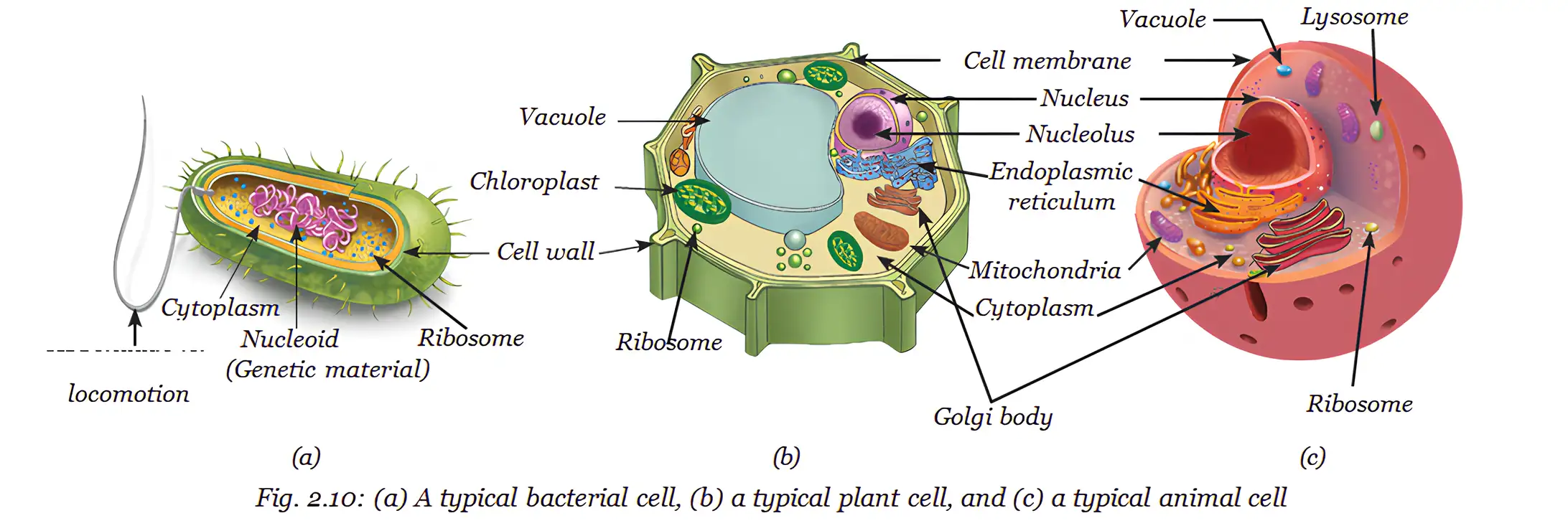
| Feature | Prokaryotic cell | Eukaryotic cell |
|---|---|---|
| Nucleus | No membrane-bound nucleus; DNA lies in a nucleoid | Well-defined, membrane-bound nucleus |
| Membrane-bound organelles | Absent | Present |
| Typical diameter | – | – |
| Organism | Usually unicellular | May be unicellular or multicellular |
| Examples | Bacteria | Plant and animal cells |
Prokaryotic cell — A cell without a well-defined, membrane-bound nucleus or membrane-bound organelles.
Eukaryotic cell — A cell with a well-defined nucleus and membrane-bound organelles.
Cell types and special contents
- Bacterial cells: Have a cell membrane, cytoplasm and cell wall; their genetic material lies in a nucleoid.
- Prokaryotic activity: Most cellular activities take place directly in the cytoplasm.
- Cytoskeleton: Fine fibres in eukaryotic cells support cell shape and help movement and internal transport; seen separately only with an electron microscope.
- Cell inclusions: Stored materials in the cytoplasm, including starch and crystals of calcium oxalate or silica in some plant cells.
- Acellular
- Not made of cells; viruses, viroids and prions are too small to be seen with a light microscope.
- Virus
- Genetic material enclosed in a protein coat.
- Viroid
- Genetic material without a protein coat.
- Prion
- A misfolded protein that has no genetic material.
Why Cells Need Organelles
Specialisation and coordination
- Division of work: Organelles let eukaryotic cells carry out different life processes independently at the same time.
- Cooperation: Organelles build materials, remove waste and provide energy, working together to keep the cell functioning.
Nucleus: The Cell’s Coded Instructions
Nuclear structures and genetic material
- Nuclear membrane: A double-layered covering with pores that allow material to pass between the nucleus and cytoplasm.
- Nucleolus: The dense region where ribosomal subunits are made; they leave the nucleus and assemble into ribosomes in the cytoplasm.
- Chromatin: Thread-like DNA-containing material in a non-dividing cell.
- Chromosomes: Before cell division, chromatin becomes organised into visible, rod-shaped chromosomes made of DNA and specific proteins.
- DNA and genes: DNA carries inherited information; functional segments of DNA are called genes.
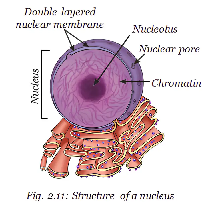
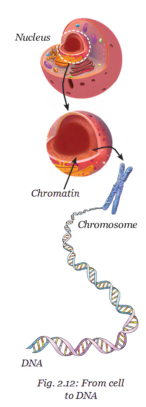
Cells without a typical nucleus
- Mature human red blood cells: Lack a nucleus, leaving more room for haemoglobin and oxygen transport. They cannot repair or divide themselves and live for about days.
- Prokaryotic cells: Their DNA is a single circular molecule associated with proteins in the nucleoid.
DNA controls cell activities
- 2010: J. Craig Venter’s team synthesised a copy of the DNA of *Mycoplasma mycoides*.
- Test: They inserted the synthetic DNA into a related bacterium after removing its DNA, while keeping the rest of its cell intact.
- Finding: The cell grew and divided under the instructions of the inserted DNA, showing that DNA controls cell structure and activities.
- Important distinction: Only the DNA was synthetic; the cell’s other parts came from an existing living cell.
Ribosomes: Protein Factories
Ribosome — A tiny structure and the site of protein synthesis.
Ribosome location
- Position: Ribosomes may lie freely in the cytoplasm or attach to the endoplasmic reticulum.
Endoplasmic Reticulum: A Manufacturing Network
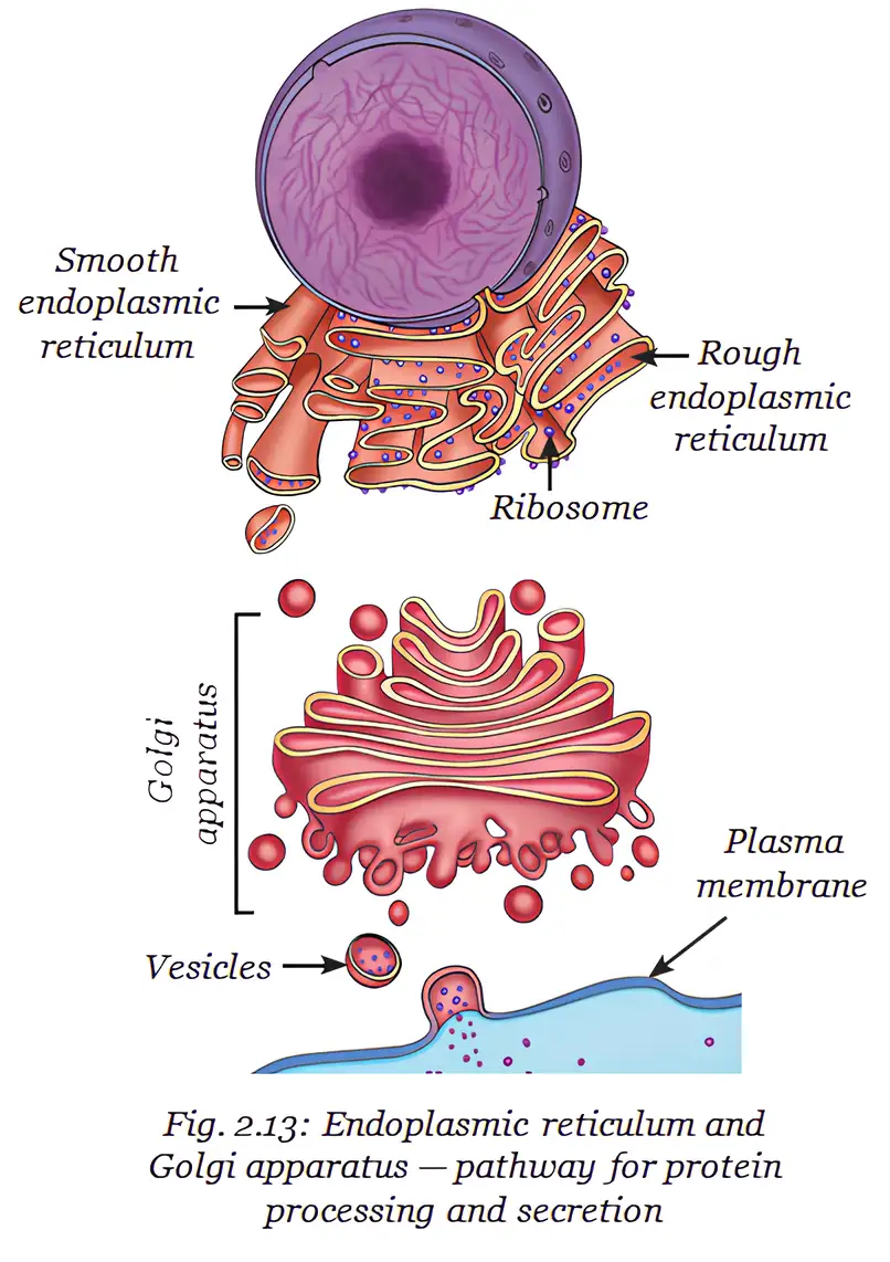
Endoplasmic reticulum (ER) — A network of membranes in the cytoplasm, continuous with the outer membrane of the nuclear envelope.
ER roles and types
- Role: Helps synthesise and transport proteins, fats (lipids) and, in some specialised cells, hormones.
- Rough ER (RER): Ribosomes on its surface give it a rough appearance; it makes proteins and helps secrete them, for example in pancreatic cells.
- Smooth ER (SER): Has no surface ribosomes; it is involved in synthesising and storing fats and hormones.
Golgi Apparatus: Packaging and Shipping Centre
Golgi apparatus — Stacks of flattened, sac-like structures that modify, sort and package cell products.
Golgi functions
- Connections: Functionally linked with the ER, cell membrane and other organelles.
- Packaging: Modifies, sorts and packages proteins and/or lipids into vesicles for transport or secretion.
- Lysosome formation: Also helps form lysosomes.
Lysosomes: The Cell’s Clean-Up System
Lysosome — A single-membrane sac containing enzymes that digest unwanted material and worn-out cell parts.
Lysosome functions
- Breakdown: Enzymes digest unwanted proteins, carbohydrates, fats and damaged cell components.
- Reuse: Breakdown products enter the cytoplasm and may be reused in other cell processes.
- Fertilisation: Lysosomal enzymes in sperm help break down the egg’s outer layer.
Mitochondria: The Cell’s Powerhouse
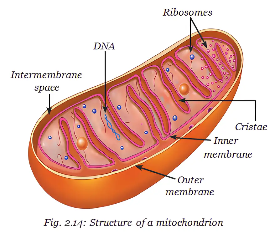
Mitochondrion — An organelle where cellular respiration releases energy from glucose and other molecules.
Structure and energy production
- Outer membrane: Smooth and porous.
- Inner membrane: Folded into finger-like projections called cristae, increasing surface area for chemical reactions and energy production.
- ATP: Energy released during respiration is stored as adenosine triphosphate (ATP), the energy currency used for most cell activities.
Plastids: Food Synthesis and Storage
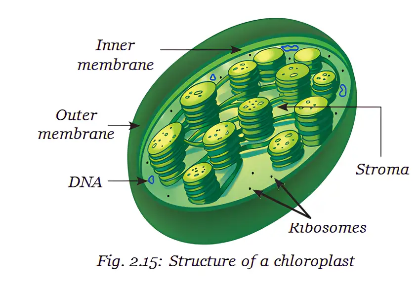
Chloroplast — A double-membrane plastid containing chlorophyll, where photosynthesis takes place.
Chloroplasts and photosynthesis
- Structure: The stroma is the semi-fluid interior; it contains disc-shaped membranes with chlorophyll.
- Photosynthesis: Chlorophyll absorbs light energy, and sugars made in photosynthesis are stored in the stroma along with starch granules.
- Shared features: Mitochondria and plastids have their own DNA and ribosomes and can make some proteins; these features suggest an evolutionary link with bacteria.
| Type | Pigment / appearance | Main role | Example |
|---|---|---|---|
| Chloroplast | Green chlorophyll | Absorbs light for photosynthesis | Green plant cells |
| Chromoplast | Coloured pigments, often yellow, orange or red | Bright colours can attract pollinators and fruit-eating animals that disperse seeds | Flower petals and fruits |
| Leucoplast | Colourless | Stores starch, oils or proteins | Starch-storing plastids in potato and taro |
White flowers may appear white because their petals reflect or scatter visible light rather than selectively absorbing it; colourless leucoplasts do not provide visible pigment.
Vacuoles: Storage and Support
Vacuole — A membrane-bound compartment for storing materials; its size and role vary between cell types.
Plant and animal vacuoles
- Mature plant cell: Usually has one large central vacuole surrounded by a selectively permeable membrane and filled with cell sap.
- Storage: Cell sap contains water, minerals, sugars and waste materials.
- Support: Water in the vacuole creates pressure that keeps the plant cell firm; water loss reduces firmness and can cause wilting.
- Animal cells: Vacuoles may be present, but are smaller and help temporarily store materials.
How do prokaryotic and eukaryotic cells differ?
How does a plant vacuole help support the cell?
How are chromatin, chromosomes and genes related?
Quick recap
- Cell types: Prokaryotes lack a membrane-bound nucleus and organelles; eukaryotes have them.
- Nucleus: DNA carries inherited information; genes are functional DNA segments, and chromatin condenses into chromosomes before division.
- Protein pathway: Ribosomes make proteins; the ER helps make and transport them, and the Golgi modifies and packages them.
- Energy and cleanup: Mitochondria release energy stored as ATP; lysosomes digest unwanted materials and worn-out parts.
- Plastids: Chloroplasts photosynthesise; chromoplasts provide colour; leucoplasts store food.
- Vacuoles: Plant vacuoles store cell sap and maintain firmness; water loss may lead to wilting.
How do Normal Cells Grow and Divide?
Cell Growth and Division
Cell division — The process by which new cells are formed from pre-existing cells.
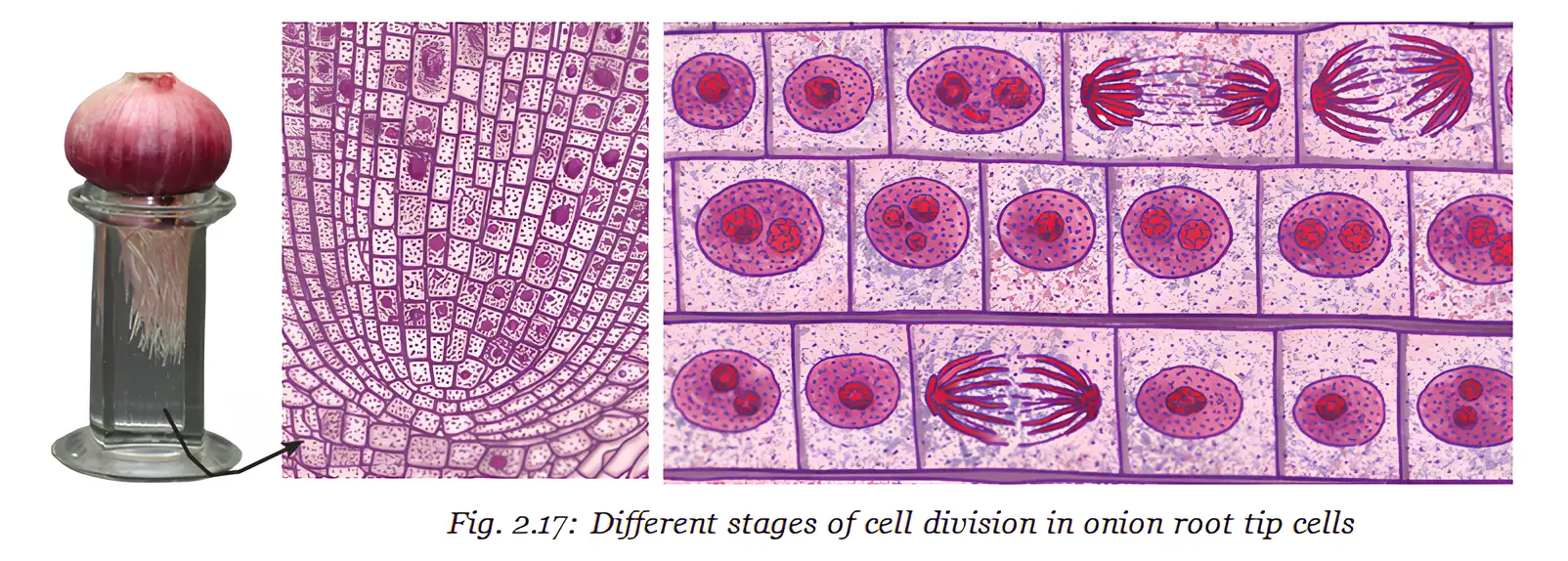
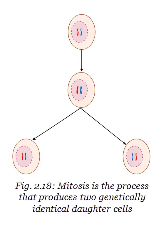
Mitosis
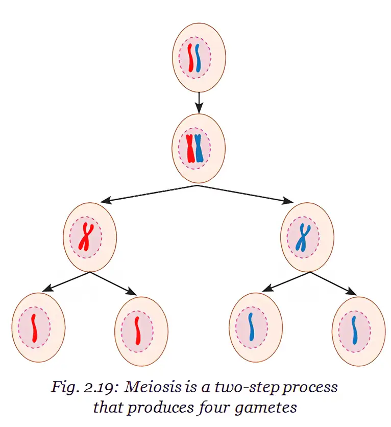
Meiosis
| Point | Mitosis | Meiosis |
|---|---|---|
| Main role | Growth, repair, maintenance and asexual reproduction | Production of gametes for sexual reproduction |
| Daughter cells formed | Two | Four |
| Chromosome number | Same as the parent cell | Half the parent cell's number |
| Genetic outcome | Daughter cells are genetically identical | Creates variation among offspring |
Cell Culture
- Cell culture: Cells are grown outside an organism in a nutrient-rich medium under sterile, carefully controlled conditions.
- Uses: Cell culture helps scientists study cells and produce biochemicals, food, medicines and vaccines.
Errors in Cell Division
How does mitosis help maintain genetic information in body cells?
How does meiosis help restore the chromosome number after fertilisation?
State one possible consequence each of errors in mitosis and meiosis.
Quick recap
- Cell division forms new cells for growth, repair and reproduction.
- Mitosis produces two genetically identical cells with the parent's DNA and chromosome number.
- Meiosis produces four gametes through two divisions, each with half the parent's DNA and chromosome number.
- Fertilisation restores the original chromosome number when two gametes combine.
- Cell culture grows cells outside the body under controlled, sterile conditions.
- Errors in mitosis or meiosis can cause problems such as tumours or genetic disorders.
Cell Theory - The Unifying Principle of Biology
Development of Cell Theory
Why Cell Theory unifies biology
- Common basis: Organisms from bacteria to humans are made of cells.
- Continuity of life: New cells form through the division of existing cells.
Totipotency and Plant Tissue Culture
- Totipotency: The ability of a living plant cell to develop into a complete plant under suitable conditions.
- Significance: Gottlieb Haberlandt proposed this idea in 1902, laying the foundation for plant tissue culture technology.
Cell Growth, Division and Death
- Cell growth: Cells grow and perform specialised functions, but growth in an organism also depends on cells dividing to form new cells.
- Cell lifespan: Cells eventually die when no longer needed, and new cells replace them to maintain tissues.
Uncontrolled Division and Cancer
- Loss of control: Errors in mitosis can cause uncontrolled cell division and tumour formation.
- Cancer: Malignant tumours can invade nearby tissues and spread to other parts of the body.
Cell Functions and Programmed Cell Death
State the three principles of classical cell theory.
What is totipotency, and why is it important?
How do contact inhibition and PCD help regulate cells?
Quick recap
- Cell theory says all organisms are cellular, cells are the basic unit of life, and cells come from existing cells.
- Schleiden (1838), Schwann (1839) and Virchow (1855) contributed to classical cell theory.
- Haberlandt proposed plant-cell totipotency in 1902, helping lay the foundation for plant tissue culture.
- Cells have definite life spans; dead cells are replaced by new cells with the same function.
- Many animal cells show contact inhibition; plant cells do not show it because of their rigid cell walls.
- Uncontrolled cell division can form tumours; cancerous tumours may invade and spread.
- PCD supports normal development, cellular quality control and immune function.
At a Glance
Cell — The basic structural and functional unit of all living organisms.
Types of Cells
| Feature | Prokaryotic cells | Eukaryotic cells |
|---|---|---|
| Nucleus | No well-defined nucleus; genetic material lies in a nucleoid | Well-defined nucleus |
| Membrane-bound organelles | Absent | Present |
| Size and complexity | Generally smaller and simpler | Generally larger and more complex |
Common Cell Structures
- Cell membrane: Surrounds every cell.
- Cell wall: Present outside the cell membrane in plant, fungal and bacterial cells.
- Cytoplasm: Fills all cells; in eukaryotic cells, it contains organelles that perform specific functions.
- Nucleus: In eukaryotic cells, it contains chromosomes made of DNA and associated proteins; chromosomes carry genetic information.
- Important organelles: Nucleus, endoplasmic reticulum, mitochondria, Golgi apparatus, ribosomes and lysosomes.
| Organelle group | Types |
|---|---|
| Plastids | Chloroplasts, leucoplasts and chromoplasts |
Cell Division
| Point | Mitosis | Meiosis |
|---|---|---|
| Process | Cell division producing daughter cells | Two-step cell division process |
| Outcome | Two daughter cells identical to the parent cell | Four daughter cells, each with half the number of chromosomes |
Normal Cells and Cancer Cells
| Point | Normal cells | Cancer cells |
|---|---|---|
| Growth and division | Grow in a controlled manner and perform their functions | Lose control and keep dividing uncontrollably |
| Outcome | Die naturally | Uncontrolled division can lead to tumour formation |
Quick recap
- The cell is the basic structural and functional unit of life.
- Prokaryotes lack a well-defined nucleus and membrane-bound organelles; eukaryotes have both.
- All cells have a cell membrane and cytoplasm; plant, fungal and bacterial cells also have a cell wall.
- Eukaryotic chromosomes contain DNA and associated proteins carrying genetic information.
- Mitosis forms two identical daughter cells; meiosis forms four cells with half the chromosome number.
- Uncontrolled cancer-cell division can form tumours.
Science — ALL CHAPTERS
1
Exploration: Entering the World of Secondary Science
2
Cell: The Building Block of Life
3
Tissues in Action
4
Describing Motion Around Us
5
Exploring Mixtures and their Separation
6
How Forces Affect Motion
7
Work, Energy, and Simple Machines
8
Journey Inside the Atom
9
Atomic Foundations of Matter
10
Sound Waves: Characteristics and Applications
11
Reproduction: How Life Continues
12
Patterns in Life: Diversity and Classification
13
Earth as a System: Energy, Matter, and Life