Class 9 . Science . Exploration . Tissues in Action
Chapter 3: Tissues in Action
Ch 3
Science
Class 9
Why are Plant and Animal Tissues Different?
From Cells to Organisms
- Organisation: Cells form tissues, tissues form organs, and organs form organ systems that make up an organism.
- Division of labour: Different tissues perform specialised functions, helping multicellular organisms carry out life processes efficiently.
Why Plant and Animal Tissues Differ
Examples of Division of Labour
- Animals: Muscle tissue enables movement; nervous tissue carries messages to different parts of the body.
- Plants: Xylem carries water and minerals, while phloem carries food.
- Cell → tissue → organ → organ system → organism.
- Tissue structure relates to its function and to the organism’s way of life.
Quick recap
- A tissue is a group of similar cells working together for a specific function.
- Multicellular organisms are organised as cells → tissues → organs → organ systems → organisms.
- Division of labour makes complex life processes more efficient.
- Plant cell walls provide rigidity and support; animal cells are more flexible, aiding movement in many animals.
- Plants make food using solar energy; animals digest food from external sources.
- Plant xylem transports water and minerals, while phloem transports food.
- Plant and animal growth patterns differ because their growth-related tissues differ in structure and function.
Tissues for Growth in Plants Part 1
Meristematic Tissue
Meristematic tissue — Plant tissue made of actively dividing cells that enables growth in length, girth and regrowth.
Main growth patterns
- Length: Roots and shoots grow longer.
- Girth: Stems increase in thickness or diameter.
- Regrowth: Plants can recover after cutting or grazing.
Apical Meristem: Growth in Length
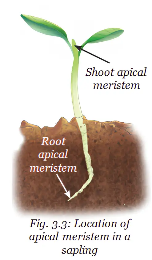
Apical meristem — Actively dividing tissue at root and shoot tips that increases the plant’s length.
Evidence and role
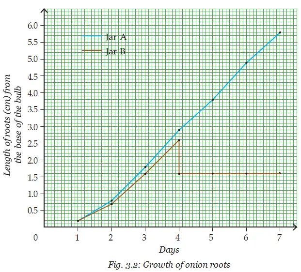
- Root tips: Roots continue growing while their tips remain intact; cutting the tips stops their growth.
- Shoot tips: Actively dividing cells at shoot tips also increase shoot length.
- Conclusion: Growth zones at root and shoot tips are called apical meristems.
Lateral Meristem: Growth in Girth
Lateral meristem — Actively dividing cells arranged around a stem that increase its diameter or girth.
How girth increases
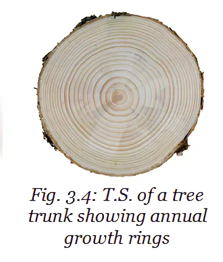
- Cell division: Lateral meristem cells form new cells towards the inside and outside in concentric layers.
- Annual growth rings: Rings visible on a tree-trunk cross-section reflect growth in different years.
- What rings indicate: Ring width reflects favourable or unfavourable conditions; counting rings can estimate a tree’s age and reveal past climatic conditions.
Intercalary Meristem: Regrowth
Intercalary meristem — Meristematic tissue at the base of an internode or just above a node that supports regrowth after cutting or grazing.
- Node
- Point on a stem from which leaves or branches arise.
- Internode
- Part of a stem between two nodes.
- Intercalary meristem
- Growth tissue at the base of an internode or just above a node.
Regrowth examples
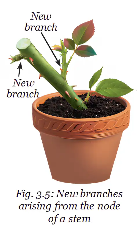
- After cutting a young stem tip: Lengthwise growth stops, but new branches can arise from stem nodes.
- Hedges: Cutting encourages new branches, making the hedge bushier.
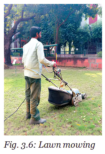
- Grass: Intercalary meristems allow grass to regrow after mowing or grazing.
| Type | Location | Main role | Example or result |
|---|---|---|---|
| Apical | Tips of roots and shoots | Increases length | Roots and shoots grow longer |
| Lateral | Along the stem circumference | Increases girth | Stem diameter increases |
| Intercalary | Base of internodes or just above nodes | Enables regrowth | Grass regrows after mowing or grazing |
Structure and Differentiation
Features of meristematic cells
- Size and walls: Cells are small and have thin cell walls.
- Cell contents: They have a large, prominent nucleus and dense cytoplasm with many organelles.
- Vacuoles: Generally absent.
- Arrangement: Cells are tightly packed, with little or no intercellular space.
- Function: These features support continuous, rapid cell division.
Differentiation — The process in which meristematic cells lose the ability to divide and become specialised in structure and function.
Formation of Permanent Tissue
- Meristematic cells divide continuously and add new cells to the plant body.
- Some new cells remain meristematic; others lose the ability to divide.
- Those cells change in structure and function through differentiation.
- They become permanent tissues specialised for functions such as support, transport or storage.
Why do roots stop growing when their tips are cut?
How do annual rings help us understand a tree?
What is differentiation, and what does it produce?
Quick recap
- Meristematic tissue consists of actively dividing cells responsible for plant growth.
- Apical meristems at root and shoot tips increase length.
- Lateral meristems around stems increase girth and produce annual growth rings.
- Intercalary meristems near nodes enable regrowth after cutting or grazing.
- Meristematic cells are small, tightly packed, and generally lack vacuoles.
- Differentiation turns some new meristematic cells into specialised permanent tissues.
Tissues for Growth in Plants Part 2
Permanent Tissues
Permanent tissue — A group of specialised plant cells that work together to perform particular functions.
Classification
- Simple permanent tissue contains one type of cell.
- Complex permanent tissue contains different types of cells that work together.
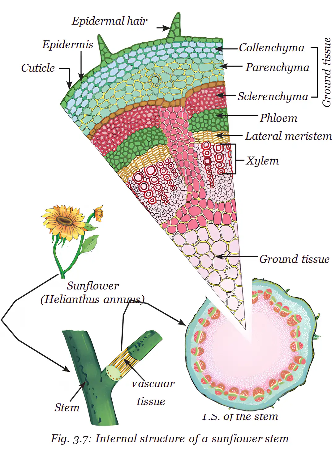
Protective Tissue: Epidermis
Epidermis — The outermost layer of the plant body, usually made of a tightly packed single layer of flat, rectangular cells.
Epidermal features and functions
- Protection: Covers plant parts and guards against mechanical injury, water loss, harmful microorganisms and harsh environmental conditions.
- Cuticle: A waxy cutin layer over epidermal cells; it limits water loss and protects against injury and parasites. It is often thicker in plants from very dry habitats.
- Root hairs: Hair-like epidermal extensions that increase surface area for absorbing water and minerals from soil.
- Stomata: Epidermal pores, especially in leaves, that allow gaseous exchange and water-vapour loss through transpiration.
- Transpiration: Water loss through stomata creates a pull in xylem that helps transport water; it also helps eliminate plant wastes.
Supporting Tissue: Simple Permanent Tissues
| Tissue | Structure | Main functions | Examples / location |
|---|---|---|---|
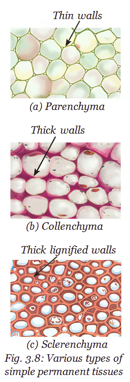
| Parenchyma | Living, thin-walled cells with intercellular spaces. | Stores food; green parenchyma can photosynthesise. In aquatic plants, air spaces aid floating. | Plant tissues; specialised air-filled parenchyma in aquatic plants. |
| Collenchyma | Living cells with unevenly thickened corners due to pectin. | Provides support and flexibility, allowing parts to bend without breaking. | Stems and tendrils. |
| Sclerenchyma | Mostly dead cells with thick, lignin-strengthened walls. | Provides hardness and strength; forms woody structures. | Stems, leaf veins, coconut husk and hard seed or nut coverings such as walnut shell. |
Conducting Tissue: Complex Permanent Tissues
| Point | Xylem | Phloem |
|---|---|---|
| Main transport | Carries water and minerals from roots to other plant parts. | Carries food from leaves to other plant parts. |
| Other function | Also provides strength to the plant. | Phloem fibres provide strength. |
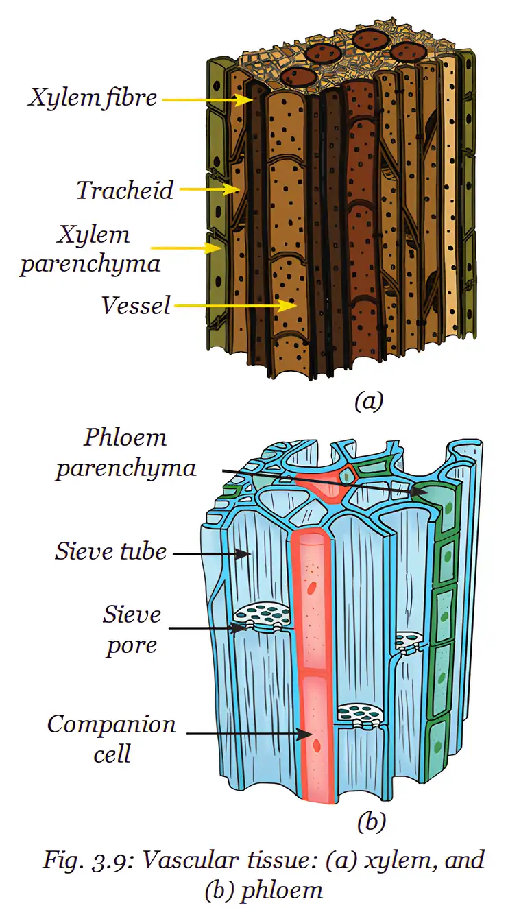
| Cell composition | Tracheids, vessels, xylem parenchyma and xylem fibres. | Sieve tubes, companion cells, phloem parenchyma and phloem fibres. |
| Living or dead | Xylem parenchyma is the only living component; the other elements are primarily sclerenchymatous. | Mostly living cells; phloem fibres are primarily sclerenchymatous. |
Roles of conducting-tissue components
- Tracheids and vessels: Thick-walled, tubular xylem cells that conduct water and minerals.
- Xylem parenchyma: Living xylem cells; the only living component of xylem.
- Sieve tubes: Long, tubular cells joined end to end by perforated walls; transport food.
- Companion cells: Specialised parenchyma cells that regulate sieve-tube functions and monitor sugar loading and unloading.
- Phloem parenchyma: Stores food, resin, tannins and latex; phloem fibres support the tissue.
Plant Tissue Systems
| Tissue system | Position / components | Main role |
|---|---|---|
| Dermal | Outer covering of the plant. | Protects inner parts and reduces water loss. |
| Ground | Main body between dermal and conducting tissues; includes parenchyma, collenchyma and sclerenchyma. | Makes up much of the plant body. |
| Vascular | Conducting tissues: xylem and phloem. | Transports water, minerals and food. |
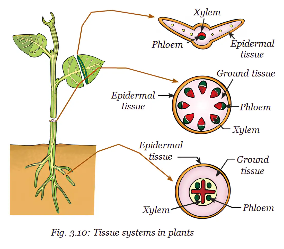
How do parenchyma, collenchyma and sclerenchyma differ?
Name the components of xylem and phloem and state their main transport roles.
Quick recap
- Permanent tissues are specialised and are simple (one cell type) or complex (multiple cell types).
- Epidermis protects the plant; cuticle reduces water loss, root hairs absorb water and minerals, and stomata enable gas exchange and transpiration.
- Parenchyma stores food, collenchyma gives flexible support, and sclerenchyma provides hardness and strength.
- Xylem transports water and minerals and supports the plant; phloem transports food.
- Companion cells regulate sieve-tube activity; phloem parenchyma stores materials and phloem fibres strengthen the tissue.
- Dermal, ground and vascular systems form the plant’s outer covering, main body and conducting tissues respectively.
Animal Tissues Part 1
Animal tissues
- Animal tissues: Groups of specialised cells work together to perform particular functions in the body.
- Main types: Animal tissues are epithelial, connective, muscular and nervous; together they provide protection, support, movement and coordination.
Epithelial tissue
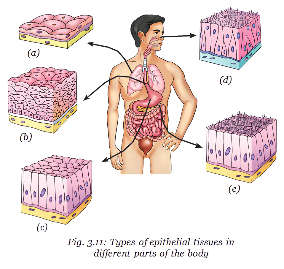
| Function | Structure | Location |
|---|---|---|
| Exchange | One layer of thin, flat cells for rapid diffusion of gases and liquids | Blood vessels and lungs |
| Protection | Many layers; outer cells are flat and tightly packed | Skin, mouth and oesophagus |
| Secretion | Cells specialised to produce and release substances; may be cuboidal or columnar | Salivary glands, sweat glands and stomach lining |
| Sensory functions | Specialised receptor cells, often with hair-like cilia | Nostrils, taste buds and inner ear |
| Absorption | One layer of tall, pillar-like cells, often with hair-like structures | Small intestine lining |
Connective tissue
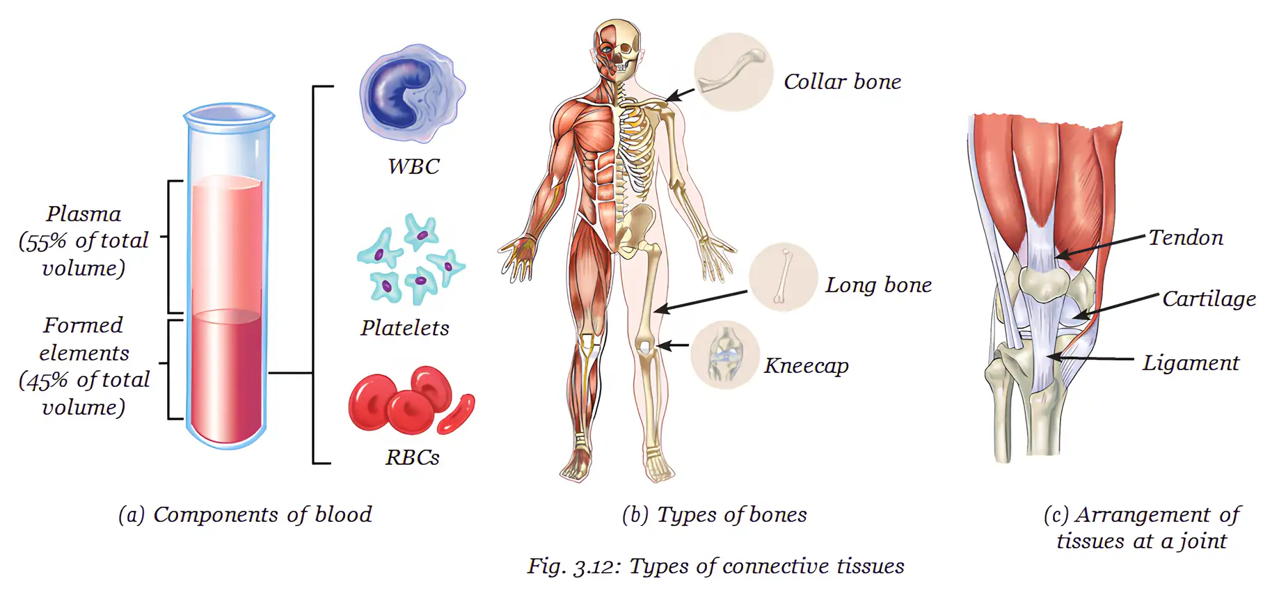
| Component or feature | Role or key fact |
|---|---|
| Red blood cells (RBCs) | Haemoglobin, an iron-rich protein, gives blood its red colour. RBCs live for about four months and are regularly replaced. |
| Platelets | Help blood clot at the site of an injury. |
| White blood cells (WBCs) | Gather at infected areas; their activity is associated with inflammation and possible pus formation. |
Connective tissue responses in everyday situations
- Injury: Platelets act at the cut → a blood clot forms.
- Infection: WBCs gather at the infected area → inflammation can cause redness and swelling, with possible pus.
- Exercise: Muscles need more oxygen → breathing speeds up and blood flow increases; the face may appear red.
| Tissue | Structure or property | Function |
|---|---|---|
| Bone | Hard, rigid matrix containing calcium and phosphorus compounds | Provides strength, support and protection |
| Cartilage | Soft, jelly-like matrix; flexible and regains its shape | Provides flexibility and cushions bone ends to absorb shock |
| Tendon | Strong connective tissue between muscle and bone | Transfers muscle action to bones to produce movement |
| Ligament | Connective tissue between bones at a joint | Stabilises joints, limits movement and helps prevent dislocation |
| Point | Tendon | Ligament |
|---|---|---|
| Connects | Muscle to bone | Bone to bone |
| Main role | Helps bring about movement | Stabilises a joint and limits excessive movement |
Do not confuse tendons with ligaments: tendons join muscle to bone; ligaments join bone to bone.
How are epithelial tissues adapted for different functions?
Differentiate between tendons and ligaments.
Quick recap
- Animal tissues are groups of similar specialised cells that perform particular functions.
- Epithelial tissue covers the body and lines organs; its structure suits exchange, protection, secretion, sensing or absorption.
- Connective tissue joins and supports body parts; its matrix gives it properties such as fluidity or rigidity.
- Haemoglobin gives RBCs their red colour; platelets help clotting, and WBCs gather at infected sites.
- Bone is hard and mineral-rich; cartilage is flexible and cushions bone ends.
- Tendons connect muscle to bone; ligaments connect bone to bone and stabilise joints.
Animal Tissues Part 2
Muscle Tissues and Movement
- Muscle function: Muscular tissue contracts to produce movement; some movements are voluntary, while others occur automatically.
- Types: Skeletal muscles move the skeleton, smooth muscles act in internal organs, and cardiac muscle contracts rhythmically in the heart.
Nervous Tissue: Control and Coordination
- Function: Nervous tissue receives, processes and transmits messages, helping control and coordinate body activities.
- Neuron: The cell body contains the nucleus, dendrites receive signals, and the axon carries messages to its terminals.
- Coordination: The brain directs responses and sends instructions to muscles and other body parts.
Quick recap
- Voluntary movements are consciously controlled; involuntary movements happen automatically.
- Skeletal muscles are unbranched, multinucleate and striated.
- Smooth muscles are spindle-shaped, single-nucleate and non-striated.
- Cardiac muscles occur only in the heart and contract rhythmically without fatigue.
- Nervous tissue coordinates body responses by receiving, processing and transmitting messages.
- A neuron has a cell body, dendrites, an axon and axon terminals.
The Musculoskeletal System
Parts and functions
Musculoskeletal system — The body system of bones, muscles, joints, cartilage, tendons and ligaments that supports the body and enables movement.
Main functions and components
- Functions: Helps us stand upright, move, maintain posture and protect delicate organs.
- Control: Its actions are controlled by the nervous system.
- Muscles and bones: Muscles pull on bones to produce movement.
- Tendons: Strong, flexible bands that attach muscles to bones.
- Body mass: In adults, the skeleton accounts for about 12–15 per cent of body weight; this varies with age, gender and body composition.
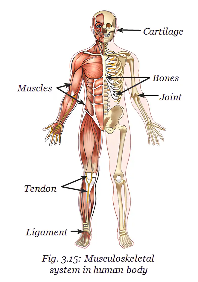
| Component | Approximate share of body weight | Note |
|---|---|---|
| Bones | 12–15 per cent | Varies with age, gender and body composition. |
| Muscles in adult males | About 40–50 per cent | An approximate range. |
| Muscles in adult females | About 30–40 per cent | An approximate range. |
How movement happens
Joint — A junction between two or more bones.
From muscle action to movement
- Joints allow bones to move relative to one another, but do not move the bones by themselves.
- Muscle contraction produces a pulling force.
- Tendon transfers the force from the contracting muscle to the bone.
- Result: The bone moves at the joint; the range and direction of movement depend on the type of joint.
Movement sequence
- Nervous system controls muscle action
- Muscle contracts and pulls
- Tendon transmits the force to the bone
- Bone moves at a joint
How do muscles produce movement at a joint?
What is a joint, and what does it do?
Quick recap
- The musculoskeletal system includes bones, muscles, joints, cartilage, tendons and ligaments.
- It supports posture and movement and protects delicate organs; the nervous system controls its actions.
- Adult bones make up about 12–15 per cent of body weight; muscle percentages vary between individuals.
- A joint is where two or more bones meet and allows movement.
- Contracting muscles pull bones through tendons, producing movement at joints.
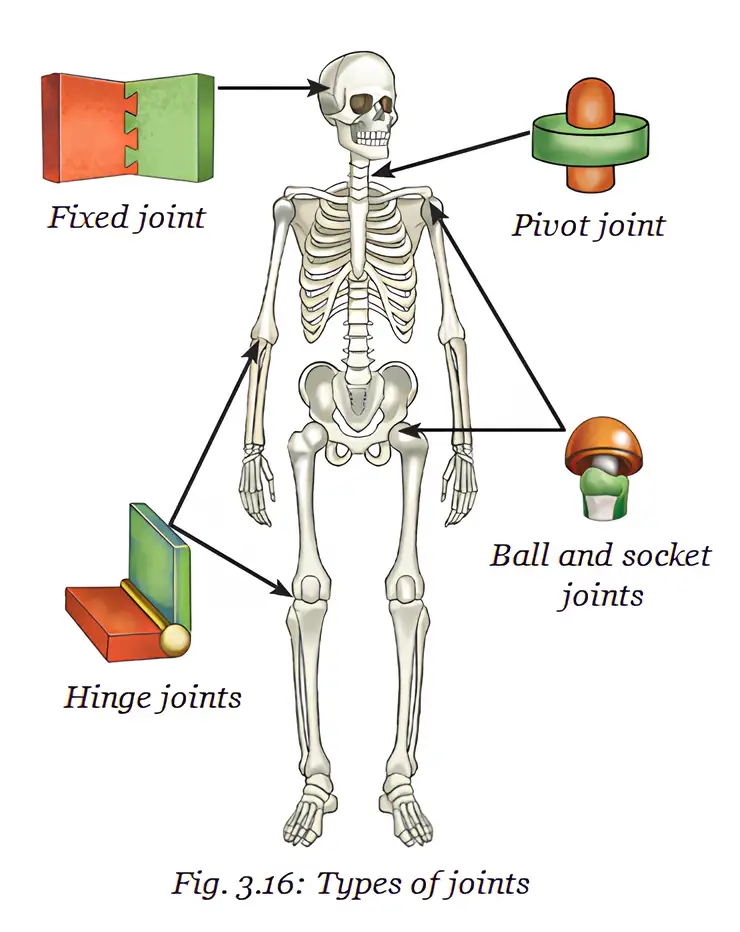
Types of Joints
Types of Joints
| Joint type | Structure or location | Movement or function |
|---|---|---|
| Ball-and-socket | Rounded top of the upper-arm bone fits into a hollow in the shoulder bone. | Moves forward, backward, sideways and in circles; example: shoulder. |
| Hinge | Found at the elbow and knee. | Bends and straightens in one direction, like a door hinge. |
| Pivot | Connects the skull to the backbone. | Allows the head to turn from side to side. |
| Fixed | Joins the flat bones of the skull. | Allows no movement; helps protect the brain, eyes and ears. |
Key details
- Shoulder girdle: The shoulder, together with the collarbone, forms the shoulder girdle, which connects the arm to the skeleton.
- Knee: The kneecap protects the knee joint.
- Skull: Fixed joints hold its bones together, keeping the protective case stable as the body moves.
Bone Marrow Stem Cells
Bone marrow stem cells — Special cells in bone marrow that can divide and produce new cells.
- Transplant use: Stem cells from a healthy donor may be given to patients with blood cancers such as leukaemia or disorders such as thalassaemia.
Quick recap
- Ball-and-socket joints allow movement in several directions, including circular movement.
- Hinge joints bend and straighten in one direction; examples are the elbow and knee.
- Pivot joints let the head turn side to side.
- Fixed joints do not move and help protect the skull’s contents.
- Bone marrow stem cells divide to make new cells and can be used in transplants.
Skeletal System
The Skeletal System
- Framework: The skeletal system is a framework of bones that supports the body and protects delicate internal organs.
- Components: It includes the skull, vertebral column and rib cage, working with muscles and joints to enable movement.
Vertebral Column and Rib Cage
- Vertebral column: A flexible backbone made of small bones called vertebrae supports the body and helps us stand upright.
- Rib cage: Twelve pairs of ribs protect the heart and lungs; flexible cartilage allows the chest to expand and contract during breathing.
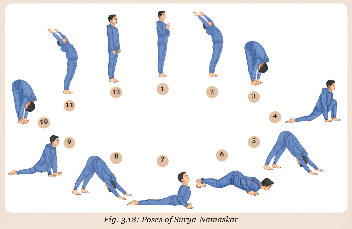
Posture, Exercise and Bone Health
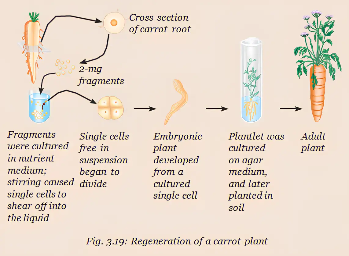
Totipotency in Plant Cells
Carrot Plant Regeneration
- Carrot phloem cell → dedifferentiation → dividing, unspecialised cell mass → redifferentiation → roots and shoots → complete plant
Carrot Cell Culture Conditions
- Culture conditions: Steward grew carrot phloem cells in nutrient medium containing simple sugars and hormones under suitable conditions.
| Light | Air | Nutrient medium | Change in fresh weight |
|---|---|---|---|
| Yes | No | Solid + nutrients | Reduced |
| Yes | Yes | Liquid + nutrients | Increased by 20% |
| No | Yes | Liquid + nutrients | Reduced |
What the Culture Results Show
- Greatest growth shown: Light, air and liquid nutrient medium together produced a 20% increase in fresh weight.
- Reduced growth: Fresh weight decreased in both the solid-medium/no-air condition and the no-light condition.
- Applications: Plant tissue culture and genetic engineering can help produce valuable phytochemicals, improved crops and disease-resistant varieties.
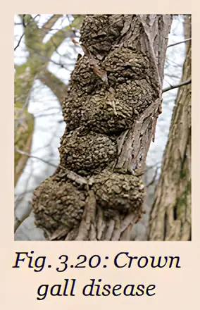
Crown Gall and Genetic Engineering
Tissues and Body Coordination
How does the rib cage help in breathing?
What is totipotency?
How is *Agrobacterium tumefaciens* useful in genetic engineering?
Quick recap
- The skeletal system supports the body and protects delicate internal organs.
- The vertebral column is made of vertebrae with cushioning cartilage discs between them.
- The 12 pairs of ribs protect the heart and lungs and move during breathing.
- Totipotency is the ability of some mature plant cells to regenerate a complete plant.
- In Steward’s culture results, light, air and liquid nutrient medium together produced a 20% increase in fresh weight.
- *Agrobacterium tumefaciens* causes crown gall and is also used to transfer useful genes into plants.
- Bones and muscles coordinate movement under nervous-system control; nervous tissue also helps regulate body activities.
Science — ALL CHAPTERS
1
Exploration: Entering the World of Secondary Science
2
Cell: The Building Block of Life
3
Tissues in Action
4
Describing Motion Around Us
5
Exploring Mixtures and their Separation
6
How Forces Affect Motion
7
Work, Energy, and Simple Machines
8
Journey Inside the Atom
9
Atomic Foundations of Matter
10
Sound Waves: Characteristics and Applications
11
Reproduction: How Life Continues
12
Patterns in Life: Diversity and Classification
13
Earth as a System: Energy, Matter, and Life