Class 11 PCB . Biology . Biology . Anatomy of Flowering Plants
Chapter 6: Anatomy Of Flowering Plants
Ch 6
Biology
Class 11 PCB
Introduction of tissue system
Plant Anatomy and Tissue Systems
Three tissue systems
- Epidermal tissue system: Forms the outer covering of the plant body.
- Ground (fundamental) tissue system: Includes tissues other than the epidermis and vascular bundles.
- Vascular (conducting) tissue system: Made of xylem and phloem.
Epidermal Tissue System
Stomatal apparatus — The stomatal pore, its two guard cells and the surrounding subsidiary cells together form the stomatal apparatus.
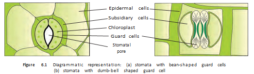
| Appendage | Structure and location | Main function |
|---|---|---|
| Root hairs | Unicellular elongations of root epidermal cells | Absorb water and minerals from soil |
| Stem trichomes | Usually multicellular; may be branched or unbranched, soft or stiff, and sometimes secretory | Help reduce water loss through transpiration |
Ground Tissue System
- Simple tissues: Ground tissue includes parenchyma, collenchyma and sclerenchyma.
- Location: Parenchyma occurs in the cortex, pericycle, pith and medullary rays; in leaves, chloroplast-containing ground tissue forms the mesophyll.
Vascular Tissue System
| Arrangement | Position of xylem and phloem | Typical occurrence |
|---|---|---|
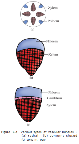
| Radial | Xylem and phloem lie alternately along different radii. | Roots |
| Conjoint | Xylem and phloem lie together along the same radius; phloem is usually outside the xylem. | Stems and leaves |
| Point | Open | Closed |
|---|---|---|
| Cambium | Present between xylem and phloem | Absent |
| Secondary tissues | Can form secondary xylem and phloem | Do not form secondary tissues |
| Typical plant group in stems | Dicotyledonous stems | Monocotyledonous stems |
What is the stomatal apparatus?
Distinguish radial and conjoint vascular bundles.
Why are some vascular bundles called open and others closed?
Quick recap
- Plant anatomy is the study of internal plant structure; cells form tissues, which form organs.
- The three tissue systems are epidermal, ground and vascular.
- The cuticle reduces water loss and is absent from roots; stomata regulate transpiration and gas exchange.
- Root hairs absorb water and minerals, while stem trichomes help reduce water loss.
- Ground tissue includes parenchyma, collenchyma and sclerenchyma; leaf ground tissue is called mesophyll.
- Vascular bundles contain xylem and phloem; open bundles have cambium, while closed bundles do not.
- Radial bundles are typical of roots; conjoint bundles are common in stems and leaves.
Anatomy Of Dicotyledonous And Monocotyledonous Plants
Studying Internal Organisation
- Transverse sections: Sections through mature roots, stems and leaves reveal how their tissues are organised.
Dicotyledonous Root
Tissue Organisation
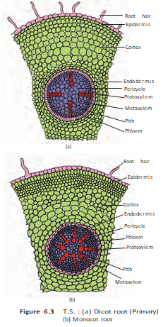
- Epiblema: Outermost layer; some cells extend outwards as unicellular root hairs.
- Cortex: Several layers of thin-walled parenchyma cells with intercellular spaces.
- Endodermis: Single innermost cortical layer of barrel-shaped cells, without intercellular spaces.
- Casparian strips: Water-impermeable suberin deposits on the radial and tangential walls of endodermal cells.
- Pericycle: A few layers of thick-walled parenchymatous cells inside the endodermis; lateral roots and vascular cambium arise from it.
- Vascular tissues: Usually 2–4 xylem and phloem patches; the parenchyma between them is conjunctive tissue.
- Pith and cambium: Pith is small or inconspicuous; a cambium ring later develops between xylem and phloem.
Stele — All tissues inside the endodermis—including the pericycle, vascular bundles and pith—form the stele.
Monocotyledonous Root
Features
- Basic organisation: Has epidermis, cortex, endodermis, pericycle, vascular bundles and pith, as in a dicot root.
- Xylem: Usually has more than six bundles; this arrangement is called polyarch.
- Pith: Large and well developed.
- Secondary growth: Does not occur in monocot roots.
| Feature | Dicot root | Monocot root |
|---|---|---|
| Xylem bundles | Usually 2–4 | Usually more than 6 (polyarch) |
| Pith | Small or inconspicuous | Large and well developed |
| Secondary growth | Can occur; a cambium ring develops | Absent |
Dicotyledonous Stem
Tissue Organisation
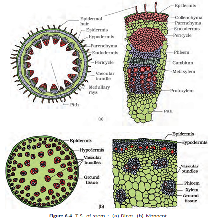
- Epidermis: Outermost protective layer, covered by a thin cuticle; it may bear trichomes and a few stomata.
- Cortex: Lies between the epidermis and pericycle and has three zones.
- Hypodermis: A few layers of collenchyma just below the epidermis; provides mechanical strength to the young stem.
- Middle cortex: Rounded, thin-walled parenchyma cells with conspicuous intercellular spaces.
- Endodermis: Innermost cortical layer; its starch-rich cells make it the starch sheath.
- Pericycle: Lies inside the endodermis and above the phloem, as semi-lunar patches of sclerenchyma.
- Medullary rays: Radially arranged parenchymatous cells between vascular bundles.
- Vascular bundles: Numerous bundles arranged in a ring; each is conjoint and open, with endarch protoxylem.
- Pith: Central region of rounded parenchyma cells with large intercellular spaces.
- Secondary growth: Occurs in most dicotyledonous stems.
Monocotyledonous Stem
Features
- Hypodermis: Sclerenchymatous.
- Ground tissue: Large, conspicuous and parenchymatous.
- Vascular bundles: Numerous and scattered; each is surrounded by a sclerenchymatous bundle sheath.
- Bundle size: Peripheral bundles are generally smaller than central bundles.
- Bundle structure: Bundles are conjoint and closed; phloem parenchyma is absent.
- Water-containing cavities: Present within the vascular bundles.
| Feature | Dicot stem | Monocot stem |
|---|---|---|
| Vascular bundles | Arranged in a ring | Scattered |
| Bundle type | Conjoint, open | Conjoint, closed |
| Hypodermis | Collenchymatous | Sclerenchymatous |
| Secondary growth | Occurs in most dicot stems | Not specified for monocot stems in this lecture |
| Other features | Starch sheath, medullary rays and central pith | Bundle sheath; phloem parenchyma absent; cavities within bundles |
Dorsiventral (Dicotyledonous) Leaf
Structure
- Main parts: Epidermis, mesophyll and vascular system.
- Epidermis: Covers both surfaces and has a conspicuous cuticle. The lower (abaxial) surface usually has more stomata; the upper (adaxial) surface may lack them.
- Mesophyll: Photosynthetic parenchyma between the epidermal layers; its cells contain chloroplasts.
- Palisade parenchyma: Elongated cells arranged vertically and parallel to one another, below the adaxial epidermis.
- Spongy parenchyma: Oval or rounded, loosely arranged cells below the palisade layer; large air spaces occur between them.
- Vascular bundles: Found in veins and the midrib; bundle size depends on vein size. In reticulate venation, veins vary in thickness.
- Bundle sheath: Each vascular bundle is surrounded by a layer of thick-walled cells.
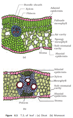
Isobilateral (Monocotyledonous) Leaf
Features
- Stomata: Present on both epidermal surfaces.
- Mesophyll: Not differentiated into palisade and spongy parenchyma.
- Bulliform cells: Large, empty, colourless adaxial epidermal cells, often found along veins in grasses.
- Leaf curling: Turgid bulliform cells expose the leaf surface; when flaccid during water stress, they curl the leaf inward and reduce water loss.
- Vascular bundles: Parallel venation is reflected in bundles of nearly similar size, except in the main veins.
| Feature | Dorsiventral (dicot) | Isobilateral (monocot) |
|---|---|---|
| Stomata | Usually more on the abaxial surface; may be absent from the adaxial surface | Present on both surfaces |
| Mesophyll | Differentiated into palisade and spongy parenchyma | Not differentiated into these two types |
| Venation and bundles | Reticulate venation; veins vary in thickness | Parallel venation; bundles are nearly similar in size except in main veins |
| Special cells | No bulliform cells specified | Bulliform cells in grasses help leaves curl during water stress |
Plant Tissues and Tissue Systems: Overview
Key Points
- Tissue types: Plant tissues are broadly meristematic (apical, lateral and intercalary) or permanent (simple and complex).
- Main functions: Tissues support food assimilation and storage, transport water, minerals and photosynthates, and provide mechanical support.
- Tissue systems: The three systems are epidermal, ground and vascular.
- Epidermal system: Includes epidermal cells, stomata and epidermal appendages.
- Ground system: Forms most of the plant body and is organised into cortex, pericycle and pith.
- Vascular system: Xylem and phloem form conducting tissues that transport water, minerals and food materials.
- Vascular bundles: Their types depend on the presence of cambium and the positions of xylem and phloem.
- Secondary growth: Occurs in most dicotyledonous roots and stems.
State three anatomical differences between dicot and monocot roots.
How do the vascular bundles of dicot and monocot stems differ?
What do bulliform cells do in grass leaves?
Quick recap
- Dicot root: Usually has 2–4 xylem patches, a small pith and the potential for secondary growth.
- Monocot root: Usually polyarch, with a large pith; it lacks secondary growth.
- Dicot stem: Vascular bundles form a ring and are conjoint and open; secondary growth occurs in most dicot stems.
- Monocot stem: Vascular bundles are scattered, conjoint and closed; secondary growth does not occur.
- Dicot leaf: Mesophyll is divided into palisade and spongy parenchyma.
- Monocot leaf: Stomata occur on both surfaces; bulliform cells can curl the leaf during water stress.
Biology — ALL CHAPTERS
1
The Living World
2
Biological Classification
3
Plant Kingdom
4
Animal Kingdom
5
Morphology of Flowering Plants
6
Anatomy Of Flowering Plants
7
Structural Organization In Animals
8
Cell The Unit Of Life
9
Biomolecules
10
Cell Cycle And Cell Division
11
Photosynthesis in Higher Plants
12
Respiration In Plants
13
Plant Growth And Development
14
Breathing And Exchange Of Gases
15
Body Fluids And Circulation
16
Excretory Products And Their Elimination
17
Locomotion And Movement
18
Neural Control And Coordination
19
Chemical Coordination and integration