Class 11 PCB . Biology . Biology . Breathing and Exchange of Gases
Chapter 14: Breathing And Exchange Of Gases
Ch 14
Biology
Class 11 PCB
The Respiratory Organs And The Mechanism Of Breathing
Breathing — The exchange of atmospheric with produced by cells; it is commonly called respiration.
Why breathing is necessary
- Oxygen supply: Cells use indirectly to break down glucose, amino acids, fatty acids and other molecules to release energy.
- Carbon dioxide removal: Catabolic reactions produce harmful , which must be removed from the body.
Respiratory Organs
- Variation: Respiratory mechanisms differ among animals, mainly with habitat and level of organisation.
- Simple diffusion: Sponges, coelenterates and flatworms exchange gases across their body surface.
- Body surface: Earthworms exchange gases through their moist cuticle.
- Tracheal tubes: Insects use a network of tubes that carries atmospheric air within the body.
| Animal group | Respiratory structure or mode | Term |
|---|---|---|
| Most aquatic arthropods and molluscs | Vascularised gills | Branchial respiration |
| Terrestrial forms | Vascularised lungs | Pulmonary respiration |
| Fishes | Gills | Branchial respiration |
| Amphibians, reptiles, birds and mammals | Lungs | Pulmonary respiration |
| Frogs | Moist skin can also exchange gases | Cutaneous respiration |
Human Respiratory System
Air pathway
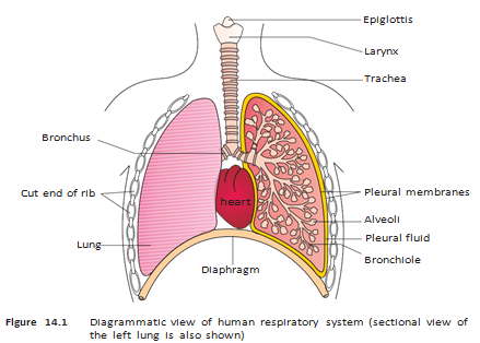
- External nostrils → nasal passage → nasal chamber
- Nasal chamber → pharynx → larynx → trachea
- Trachea → right and left primary bronchi → secondary and tertiary bronchi
- Tertiary bronchi → bronchioles → terminal bronchioles → alveoli
Important airway structures
- Pharynx: A shared passage for food and air.
- Larynx: A cartilaginous sound box that helps produce sound.
- Epiglottis: A thin, elastic cartilage flap that covers the glottis during swallowing and helps prevent food entering the larynx.
- Trachea: A straight tube reaching the mid-thoracic cavity; it divides into two primary bronchi at the level of the fifth thoracic vertebra.
- Cartilage support: The trachea, primary, secondary and tertiary bronchi, and initial bronchioles have incomplete cartilaginous rings.
- Alveoli: Thin-walled, irregular, vascularised air sacs arising from terminal bronchioles; they are the main sites of gas exchange.
- Lungs: The branching bronchi, bronchioles and alveoli together form the lungs.
Pleura and respiratory parts
- Pleura: Each lung is covered by two pleural membranes, with pleural fluid between them to reduce friction.
- Membrane positions: The outer pleural membrane contacts the thoracic lining; the inner membrane contacts the lung surface.
- Conducting part: Extends from the external nostrils to the terminal bronchioles; transports air, removes foreign particles, humidifies it and brings it to body temperature.
- Exchange part: Consists of alveoli and their ducts; and diffuse between alveolar air and blood.
Thoracic chamber
- Structure: The lungs lie in an anatomically airtight thoracic chamber.
- Boundaries: Vertebral column behind, sternum in front, ribs at the sides and dome-shaped diaphragm below.
- Volume connection: Changes in thoracic-cavity volume are reflected in lung-cavity volume; this arrangement enables breathing because lung volume cannot be altered directly.
Steps of respiration
- Pulmonary ventilation: atmospheric air enters and -rich alveolar air leaves.
- Diffusion: and move across the alveolar membrane.
- Blood transports the gases.
- and diffuse between blood and tissues.
- Cells use for catabolic reactions and release during cellular respiration.
What are the conducting and exchange parts of the human respiratory system?
Quick recap
- Breathing supplies cells with and removes the they produce.
- Animal respiratory structures include body surfaces, tracheal tubes, gills and lungs.
- In humans, air travels from the nostrils through branching airways to the alveoli.
- The larynx produces sound; the epiglottis helps keep swallowed food out of the larynx.
- Pleural fluid reduces friction, while thoracic-cavity volume changes drive lung-volume changes.
- Respiration includes ventilation, gas diffusion, blood transport, tissue exchange and cellular use of oxygen.
Mechanism Of Breathing
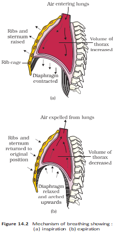
Breathing — The movement of air into and out of the lungs occurs in two stages: inspiration and expiration.
| Feature | Inspiration | Expiration |
|---|---|---|
| Muscle action | Diaphragm contracts; external intercostals lift the ribs and sternum. | Diaphragm and intercostal muscles relax; diaphragm and sternum return to normal positions. |
| Thoracic volume | Increases: diaphragm contraction increases the anteroposterior axis; external intercostals lift the ribs and sternum to increase the dorsoventral axis. | Decreases as the muscles relax. |
| Pulmonary volume | Increases | Decreases |
| Intrapulmonary pressure | Falls below atmospheric pressure | Rises slightly above atmospheric pressure |
| Air movement | Atmosphere → lungs | Lungs → outside |
Pressure changes during breathing
- Inspiration: Diaphragm contracts and external intercostals lift the ribs and sternum → thoracic volume increases in the anteroposterior and dorsoventral axes → pulmonary volume increases → intrapulmonary pressure falls below atmospheric pressure → air enters.
- Expiration: Diaphragm and intercostal muscles relax → thoracic and pulmonary volumes decrease → intrapulmonary pressure rises above atmospheric pressure → air is expelled.
Breathing rate and measurement
- Additional muscles: Abdominal muscles can increase the strength of inspiration and expiration.
- Breathing rate: A healthy human breathes about 12–16 times per minute.
- Spirometer: Measures air volumes during breathing and helps assess pulmonary function clinically.
Respiratory Volumes and Capacities
Respiratory volumes
- Tidal volume (TV): Air inspired or expired during normal breathing; about 500 mL. This corresponds to about 6,000–8,000 mL per minute.
- Inspiratory reserve volume (IRV): Additional air that can be forcibly inspired; about 2,500–3,000 mL.
- Expiratory reserve volume (ERV): Additional air that can be forcibly expired; about 1,000–1,100 mL.
- Residual volume (RV): Air remaining in the lungs after forcible expiration; about 1,100–1,200 mL.
| Capacity | Definition | Components |
|---|---|---|
| Inspiratory capacity (IC) | Total air inspired after a normal expiration. | |
| Expiratory capacity (EC) | Total air expired after a normal inspiration. | |
| Functional residual capacity (FRC) | Air remaining in the lungs after a normal expiration. | |
| Vital capacity (VC) | Maximum air breathed in after a forced expiration, or breathed out after a forced inspiration. | |
| Total lung capacity (TLC) | Total air in the lungs at the end of a forced inspiration. |
Exchange of Gases
Gas exchange by diffusion
- Sites: Alveoli are the primary gas-exchange sites; gases are also exchanged between blood and body tissues.
- Mechanism: Oxygen and carbon dioxide move by simple diffusion, mainly down their partial-pressure gradients.
- Other factors: Gas solubility and diffusion-membrane thickness also affect the rate of diffusion.
- Partial pressure: The pressure contributed by an individual gas in a mixture; oxygen and carbon dioxide partial pressures are written as and .
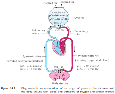
| Gas | Atmospheric air | Alveoli | Deoxygenated blood | Oxygenated blood | Tissues |
|---|---|---|---|---|---|
| 159 | 104 | 40 | 95 | 40 | |
| 0.3 | 40 | 45 | 40 | 45 |
Gas diffusion gradients
- Oxygen: Alveoli ( mm Hg) → deoxygenated blood ( mm Hg); then oxygenated blood ( mm Hg) → tissues ( mm Hg).
- Carbon dioxide: Tissues ( mm Hg) → oxygenated blood ( mm Hg); then deoxygenated blood ( mm Hg) → alveoli ( mm Hg).
Diffusion membrane and factors
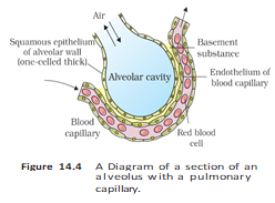
- Layers: The membrane includes thin squamous alveolar epithelium, the alveolar-capillary endothelium and the basement substance between them.
- Basement substance: It comprises the thin basement membrane supporting the alveolar epithelium and the basement membrane around the single-layer capillary endothelium.
- Thickness: The complete diffusion membrane is much less than 1 mm thick.
- Solubility: is 20–25 times more soluble than ; therefore, more diffuses per unit difference in partial pressure.
How do inspiration and expiration create airflow?
State the components of vital capacity and total lung capacity.
Quick recap
- Breathing has two stages: inspiration and expiration, driven by pressure differences between lungs and atmosphere.
- During inspiration, diaphragm contraction increases anteroposterior volume; external intercostals lift the ribs and sternum to increase dorsoventral volume.
- Inspiration increases thoracic volume and lowers intrapulmonary pressure; expiration reverses these changes.
- TV, IRV, ERV and RV are the four respiratory volumes; pulmonary capacities are combinations of them.
- Alveoli and tissues exchange gases with blood by diffusion down partial-pressure gradients.
- moves from alveoli to blood and then tissues; moves from tissues to blood and then alveoli.
- High solubility and a very thin diffusion membrane favour gas exchange.
Transport Of Gases
How Gases Are Transported in Blood
Blood as the transport medium
- Blood transports both and between the lungs and body tissues.
- Oxygen: About is carried by red blood cells (RBCs); the remaining is dissolved in plasma.
- Carbon dioxide: About – is carried by RBCs, about as bicarbonate, and about dissolved in plasma.
| Gas | Main transport form | Other transport form |
|---|---|---|
| About carried by RBCs, bound to haemoglobin | About dissolved in plasma | |
| About as bicarbonate; – carried by RBCs | About dissolved in plasma |
Transport of Oxygen
Haemoglobin — A red-coloured, iron-containing pigment in RBCs that binds oxygen reversibly to form oxyhaemoglobin.
Oxygen binding and release
- Capacity: Each haemoglobin molecule can carry up to four molecules.
- Main factor: Binding depends primarily on the partial pressure of oxygen, .
- Other influences: , hydrogen-ion concentration () and temperature can alter oxygen binding.
- Oxygen dissociation curve: A sigmoid curve showing haemoglobin's percentage saturation with as changes; it helps explain how these factors affect binding.
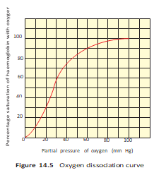
| Condition | Alveoli | Tissues |
|---|---|---|
| High | Low | |
| and | Low | High |
| Temperature | Lower | Higher |
| Effect on haemoglobin | Conditions favour oxyhaemoglobin formation and binding | Conditions favour dissociation and delivery to tissues |
- Under normal conditions, of oxygenated blood delivers about of to tissues.
Transport of Carbon Dioxide
Carbamino-haemoglobin transport
- Carbamino-haemoglobin: About – of is carried by binding to haemoglobin.
- Binding factors: The binding depends on and is strongly affected by .
- At tissues: High and low promote binding to haemoglobin.
- At alveoli: Low and high promote dissociation, releasing it for exhalation.
Bicarbonate transport
- Carbonic anhydrase: This enzyme is present in high concentration in RBCs and in minute amounts in plasma; it speeds the reaction in both directions.
- At tissues: High from cellular catabolism causes to enter blood and form bicarbonate () and .
- At alveoli: Low shifts the reaction in reverse, forming and water; the is then released.
- Net delivery: Bicarbonate carries much of the tissue-produced to the alveoli.
- Every of deoxygenated blood delivers about of to the alveoli.
Regulation of Respiration
| Structure | Location | Role |
|---|---|---|
| Respiratory rhythm centre | Medulla | Primarily generates and regulates the respiratory rhythm. |
| Pneumotaxic centre | Pons | Moderates the rhythm centre; neural signals can shorten inspiration and alter respiratory rate. |
| Chemosensitive area | Adjacent to the rhythm centre | Responds strongly to increases in and ; signals the rhythm centre to adjust breathing and remove them. |
| Receptors | Aortic arch and carotid artery | Detect changes in and and signal the rhythm centre for corrective action. |
Key regulatory principle
- Control: The neural system adjusts respiratory rhythm to meet the needs of body tissues.
- Oxygen: Its role in regulating respiratory rhythm is quite insignificant compared with the effects of and .
How do conditions in the alveoli and tissues affect oxygen binding to haemoglobin?
Describe the role of the respiratory rhythm centre, pneumotaxic centre and chemosensitive area.
Quick recap
- Blood transports mainly through RBC haemoglobin and transports most as bicarbonate.
- Each haemoglobin molecule can bind up to four oxygen molecules; oxygen binding varies with , , and temperature.
- Alveolar conditions favour oxygen loading; tissue conditions favour oxygen unloading.
- Carbon dioxide travels partly as carbamino-haemoglobin and mainly as bicarbonate formed with carbonic anhydrase.
- The medullary rhythm centre regulates breathing, while the pons, chemosensitive area and arterial receptors modify it.
- Changes in and strongly influence respiratory regulation; oxygen's role is insignificant.
Disorders Of Respiratory System
Asthma
- Asthma: Inflammation of the bronchi and bronchioles causes difficulty in breathing and wheezing.
Emphysema
- Emphysema: This chronic disorder damages alveolar walls, reducing the respiratory surface; cigarette smoking is a major cause.
Occupational Respiratory Disorders
- Occupational exposure: Prolonged exposure to dust in industries such as grinding and stone-breaking can inflame the lungs and cause fibrosis.
- Prevention: Workers in dusty industries should wear protective masks.
Respiration: Main Stages
- Respiration: It includes breathing, gas exchange between alveoli and blood, transport of gases, exchange between blood and tissues, and cellular use of oxygen.
Breathing Mechanics and Measurement
- Breathing: Inspiration draws air into the lungs, while expiration releases alveolar air; changes in thoracic volume create the pressure gradients that move air.
- Measurement: A spirometer measures respiratory volumes and capacities and helps assess pulmonary function.
Diffusion of Respiratory Gases
- Gas exchange: Oxygen and carbon dioxide diffuse across alveolar and tissue surfaces down their partial-pressure gradients.
- Diffusion factors: Gas solubility and the thickness of the diffusion membrane also affect the rate of exchange.
Transport of Oxygen and Carbon Dioxide
- Oxygen: Most oxygen is carried by haemoglobin in red blood cells as oxyhaemoglobin; a small amount is dissolved in plasma.
- Carbon dioxide: It is transported mainly as bicarbonate, with the remainder carried by haemoglobin or dissolved in plasma.
Control of Respiratory Rhythm
What is emphysema, and what is a major cause?
How are oxygen and carbon dioxide transported in blood?
How can prolonged exposure to industrial dust damage the lungs?
Quick recap
- Asthma inflames the bronchi and bronchioles, causing breathing difficulty and wheezing; emphysema damages alveolar walls.
- Prolonged industrial dust exposure can cause inflammation, fibrosis and serious lung damage; protective masks are recommended.
- Respiration includes breathing, gas exchange at alveoli and tissues, blood transport, and cellular use of oxygen.
- Intercostal muscles and the diaphragm create pressure gradients for breathing; a spirometer estimates breathing-related air volumes.
- Diffusion depends on partial-pressure gradients, solubility and diffusion-surface thickness; oxygen moves towards tissues and carbon dioxide towards alveoli.
- Oxygen is carried mainly as oxyhaemoglobin; carbon dioxide is carried chiefly as bicarbonate and carbamino-haemoglobin.
- The medulla respiratory centre maintains rhythm; the pons pneumotaxic centre and medullary chemosensitive area can alter breathing.
Biology — ALL CHAPTERS
1
The Living World
2
Biological Classification
3
Plant Kingdom
4
Animal Kingdom
5
Morphology of Flowering Plants
6
Anatomy Of Flowering Plants
7
Structural Organization In Animals
8
Cell The Unit Of Life
9
Biomolecules
10
Cell Cycle And Cell Division
11
Photosynthesis in Higher Plants
12
Respiration In Plants
13
Plant Growth And Development
14
Breathing And Exchange Of Gases
15
Body Fluids And Circulation
16
Excretory Products And Their Elimination
17
Locomotion And Movement
18
Neural Control And Coordination
19
Chemical Coordination and integration