Class 11 PCB . Biology . Biology . Cell the Unit of Life
Chapter 8: Cell The Unit Of Life
Ch 8
Biology
Class 11 PCB
What Is Cell?
The Cell: Basic Unit of Life
- Cell: The fundamental structural and functional unit of all living organisms.
- Living organisms: Composed of one or more cells; single-celled organisms can independently perform essential life functions.
Cell Theory
Development of Cell Theory
- 1838Matthias Schleiden concluded from studying plants that they are composed of different kinds of cells forming tissues.
- 1839Theodor Schwann studied animal cells, described their thin outer layer (now called the plasma membrane), and noted that plant cells have a cell wall.
- 1855Rudolf Virchow stated that cells divide and new cells arise from pre-existing cells: Omnis cellula e cellula.
Cell Theory: Main Ideas
- Schleiden and Schwann formulated the cell theory: plants and animals are composed of cells and cell products.
- Virchow added that new cells form by division of pre-existing cells, completing the theory.
- All living organisms are composed of cells and cell products.
- All cells arise from pre-existing cells.
Overview of Cell Structure
Basic Cell Components
- Cell boundary: Plant cells have an outer cell wall with a cell membrane just inside it; cheek cells are bounded by a cell membrane.
- Nucleus: A dense, membrane-bound structure containing chromosomes; chromosomes contain DNA, the genetic material.
- Cytoplasm: A semi-fluid matrix filling the cell; it is the main site of cellular activities and chemical reactions that maintain life.
| Feature | Prokaryotic cells | Eukaryotic cells |
|---|---|---|
| Nucleus | Lack a membrane-bound nucleus | Have a membrane-bound nucleus |
| Membrane-bound organelles | Absent | Present, including ER, Golgi complex, lysosomes, mitochondria, microbodies and vacuoles |
| Ribosomes | Present; not membrane-bound | Present; not membrane-bound |
Organelles and Their Locations
- Ribosomes occur in the cytoplasm of both cell types; in eukaryotic cells they are also found in mitochondria, chloroplasts and on rough ER.
- Chloroplasts occur in plant cells; mitochondria and chloroplasts contain ribosomes.
- Centrosome: An animal-cell organelle without a membrane; it helps in cell division.
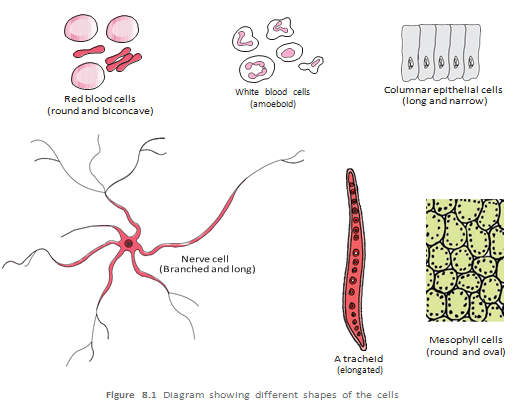
Cell Size, Shape and Function
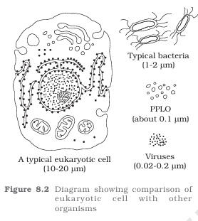
- Size range: Mycoplasmas are among the smallest cells, about 0.3 µm long; bacteria may be 3–5 µm.
- Largest isolated single cell: The egg of an ostrich.
- Human red blood cells are about 7.0 µm in diameter; nerve cells are among the longest cells.
- Shapes include disc-like, polygonal, columnar, cuboid, thread-like and irregular forms.
- Shape and function: Cell shape varies according to the function the cell performs.
State the two statements of cell theory.
Give two differences between prokaryotic and eukaryotic cells.
Quick recap
- The cell is the basic structural and functional unit of life.
- Unicellular organisms live independently; multicellular organisms contain many cells.
- Schleiden and Schwann formulated cell theory; Virchow added that cells arise from pre-existing cells.
- Eukaryotic cells have a membrane-bound nucleus and organelles; prokaryotic cells lack both.
- Ribosomes are non-membrane-bound and occur in both prokaryotic and eukaryotic cells.
- Cells vary in size and shape, often in relation to their function.
Prokaryotic Cell
General Features and Shapes
Prokaryotic cell — A cell without a well-defined nucleus enclosed by a nuclear membrane.
- Examples: Bacteria, blue-green algae (cyanobacteria), mycoplasma and PPLO (Pleuro Pneumonia Like Organisms).
- Size and growth: Prokaryotic cells are generally smaller and multiply more rapidly than eukaryotic cells.
- Genetic material: Usually a single, circular genomic DNA molecule that is naked (not enclosed by a nuclear membrane); eukaryotic cells have a nuclear membrane.
- Plasmids: Many bacteria also have small circular DNA molecules outside the genomic DNA; plasmids can confer traits such as antibiotic resistance.
- Application: Plasmid DNA can be used to monitor bacterial transformation with foreign DNA.
- Cell contents: The cytoplasm is the fluid matrix filling the cell. Eukaryote-like organelles are absent except for ribosomes; inclusions are also present.
| Bacterial shape | Description |
|---|---|
| Bacillus | Rod-shaped |
| Coccus | Spherical |
| Vibrio | Comma-shaped |
| Spirillum | Spiral-shaped |
Cell Envelope and Its Modifications
Cell envelope — A protective, three-layered covering found in most prokaryotes, especially bacteria.
- Layers, outside to inside: Glycocalyx → cell wall → plasma membrane; together they act as one protective unit.
- Glycocalyx: Its composition and thickness vary; it may be a loose slime layer or a thick, tough capsule.
- Cell wall: Determines cell shape and provides structural support, helping prevent the bacterium from bursting or collapsing.
- Plasma membrane: Selectively permeable, interacts with the surroundings and is structurally similar to the eukaryotic plasma membrane.
- Mycoplasma: Lacks a cell wall.
| Feature | Gram-positive | Gram-negative |
|---|---|---|
| Response to Gram staining | Takes up the Gram stain | Does not take up the Gram stain |
| Basis of grouping | Differences in cell envelopes and staining response | Differences in cell envelopes and staining response |
Mesosome — An infolding of the plasma membrane into the cell, forming vesicles, tubules or lamellae.
Mesosome Functions
- Functions: Helps in cell-wall formation, DNA replication and distribution to daughter cells, respiration and secretion.
- Additional roles: Increases the plasma membrane’s surface area and enzymatic content.
Chromatophores — Pigment-containing membranous extensions into the cytoplasm found in some prokaryotes, such as cyanobacteria.
| Structure | Description | Main role |
|---|---|---|
| Flagella | Thin, filamentous extensions from the cell wall; present in motile bacteria | Enable motility; number and arrangement vary. Each has a filament, hook and basal body. |
| Pili | Elongated tubular structures made of a special protein | Do not play a role in motility. |
| Fimbriae | Small, bristle-like fibres projecting from the cell | Do not enable motility; some help bacteria attach to rocks in streams or to host tissues. |
- Flagellum parts: Filament → hook → basal body; the filament is the longest part and extends from the cell surface to the outside.
Ribosomes and Inclusion Bodies
Prokaryotic ribosome — A protein-synthesising particle associated with the plasma membrane in prokaryotes.
- Size: About .
- Subunits: The and subunits together form a ribosome.
- Polysome: Several ribosomes attached to one mRNA form a polysome (polyribosome); each ribosome translates the mRNA into protein.
Inclusion bodies — Non-membrane-bound deposits in the cytoplasm that store reserve materials in prokaryotic cells.
- Examples: Phosphate granules, cyanophycean granules and glycogen granules.
- Gas vacuoles: Found in blue-green, purple and green photosynthetic bacteria.
Describe the bacterial cell envelope and the roles of its layers.
What are prokaryotic ribosomes and polysomes?
Quick recap
- Prokaryotes are generally small, multiply rapidly and lack a well-defined, membrane-bound nucleus.
- The fluid matrix filling a prokaryotic cell is the cytoplasm; bacteria also have genomic DNA and may contain plasmids.
- Bacteria have four common shapes: bacillus, coccus, vibrio and spirillum.
- The envelope is glycocalyx → cell wall → plasma membrane; glycocalyx varies, and mycoplasma lacks a cell wall.
- Mesosomes are plasma-membrane infoldings; chromatophores contain pigments in some prokaryotes.
- Flagella enable motility and vary in number and arrangement; pili and fimbriae do not enable motility.
- Prokaryotic ribosomes are , formed from and subunits; inclusion bodies store reserves.
Eukaryotic Cells
Eukaryotic Cells
- Eukaryotes: Protists, plants, animals and fungi; their cells have an organised, membrane-bound nucleus and membrane-bound organelles.
- Plant and animal cells: Plant cells have cell walls, plastids and a large central vacuole; animal cells have centrioles, usually absent in plants.
Cell Membrane
Fluid Mosaic Model and Membrane Function
- Model: Singer and Nicolson proposed the fluid mosaic model in 1972.
- Fluidity: The quasi-fluid lipid bilayer allows lateral movement of proteins within the membrane.
- Importance: Membrane fluidity supports cell growth, intercellular junction formation, secretion, endocytosis and cell division.
- Selectively permeable: Only certain molecules can cross the membrane.
Cell Wall
- Structure: A non-living, rigid covering outside the plasma membrane in plants and fungi.
- Functions: Gives shape, protects against mechanical damage and infection, and helps in cell-to-cell interaction.
- Plant cell wall: The primary wall can grow; a secondary wall may form inside it as the cell matures. The calcium-pectate middle lamella joins neighbouring cells.
Endomembrane System
- Components: The endomembrane system comprises the endoplasmic reticulum, Golgi complex, lysosomes and vacuoles.
- Coordination: These organelles work together in related cellular functions; mitochondria, chloroplasts and peroxisomes are not included because their functions are not coordinated with this system.
Endoplasmic Reticulum (ER)
- Structure: The endoplasmic reticulum is a network of tiny tubules that separates the cell interior into luminal and cytoplasmic compartments.
- Rough ER: Ribosome-covered and prominent in cells involved in protein synthesis and secretion.
- Smooth ER: Lacks ribosomes and is a major site of lipid synthesis; in animal cells it also synthesises steroid hormones.
Golgi Apparatus
- Structure: The Golgi apparatus consists of stacks of flattened, disc-shaped cisternae with distinct cis (forming) and trans (maturing) faces.
- Function: It modifies and packages materials for delivery within the cell or secretion outside it.
- Products: It is an important site for the formation of glycoproteins and glycolipids.
Lysosomes
- Structure: Lysosomes are membrane-bound vesicles formed by packaging in the Golgi apparatus.
- Enzymes: They contain hydrolytic enzymes, including lipases, proteases and carbohydrases, that work best at acidic pH.
- Function: Their enzymes digest carbohydrates, proteins, lipids and nucleic acids.
Vacuoles
Differentiate between passive and active transport across the plasma membrane.
What is the difference between RER and SER?
Name the endomembrane system components and two organelles excluded from it.
Quick recap
- Eukaryotic cells have an organised nucleus, chromosome-based genetic material and membrane-bound organelles.
- Plant and animal cells differ in cell walls, plastids, central vacuoles and centrioles.
- The fluid mosaic membrane is selectively permeable; transport may be passive, facilitated or ATP-dependent.
- Cell walls protect and support cells; plasmodesmata connect neighbouring plant-cell cytoplasms.
- The endomembrane system consists of the ER, Golgi complex, lysosomes and vacuoles.
- RER supports protein production, SER synthesises lipids, and the Golgi modifies and packages materials.
- Lysosomes digest cell materials; vacuoles store substances and have specialised roles in different cells.
Mitochondria
Mitochondria
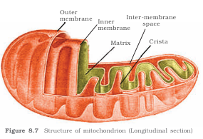
Mitochondrion — A double-membrane organelle and the main site of aerobic respiration and ATP production in eukaryotic cells.
- Visibility and number: Mitochondria are not easily seen without specific staining; their number varies with the cell’s physiological activity.
- Shape and size: Commonly sausage-shaped or cylindrical; diameter is – (average ) and length is –.
- Compartments: The outer membrane forms the boundary; the inner membrane divides the lumen into outer and inner compartments.
- Cristae: Infoldings of the inner membrane that increase surface area; both membranes have enzymes involved in mitochondrial functions.
- Matrix: Contains a circular DNA molecule, some RNA, ribosomes and components for protein synthesis.
- Division and role: Mitochondria divide by fission and produce ATP during aerobic respiration; hence, they are called the cell’s “powerhouses.”
Plastids
Plastid — A large organelle found in plant cells and euglenoids; plastids may contain pigments or store nutrients.
| Type | Pigment or contents | Main role / example |
|---|---|---|
| Chloroplast | Chlorophyll and carotenoids | Traps light energy for photosynthesis |
| Chromoplast | Fat-soluble carotenoids, including carotene and xanthophylls | Colours plant parts yellow, orange or red |
| Leucoplast | Colourless; stores nutrients | Amyloplasts store starch (e.g. potato); elaioplasts store oils and fats; aleuroplasts store proteins |
Chloroplast structure and functions
- Location: Most chloroplasts in green plants occur in the leaf mesophyll.
- Shape and size: Lens-shaped, oval, spherical, discoid or ribbon-like; typically – long and – wide.
- Number: From one per *Chlamydomonas* cell to about – per mesophyll cell.
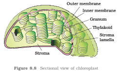
- Envelope and stroma: A double membrane surrounds the chloroplast; the inner membrane is less permeable. The enclosed space is the stroma.
- Thylakoids and grana: Flattened thylakoid sacs occur in the stroma; stacks are grana (singular: granum). Unstacked connecting thylakoids are intergranal thylakoids, forming stroma lamellae between grana.
- Lumen: Each thylakoid encloses an internal space called the lumen.
- Contents: Thylakoid membranes contain chlorophyll; the stroma contains enzymes for carbohydrate and protein synthesis, circular DNA and ribosomes.
- Photosynthesis: Light reactions occur in the grana, while dark reactions occur in the stroma.
- Ribosomes: Chloroplast ribosomes are , smaller than cytoplasmic ribosomes.
Ribosomes
Ribosome — A non-membrane-bound structure made of RNA and proteins that carries out protein synthesis.
- Discovery: George Palade first observed ribosomes as dense particles under an electron microscope in 1953.
- Structure: Each ribosome has a larger and a smaller subunit.
- Svedberg unit: denotes the sedimentation coefficient, an indirect measure of particle size and density.
| Ribosome | Large subunit | Small subunit |
|---|---|---|
| Eukaryotic | ||
| Prokaryotic |
Do not add the subunit values to determine the ribosome’s value: is a sedimentation coefficient, not an arithmetic total.
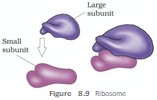
Cytoskeleton
Cytoskeleton — A network of protein filaments in the cytoplasm that supports the cell and helps it maintain shape and move.
- Components: Microtubules, microfilaments and intermediate filaments.
- Functions: Mechanical support, cell motility and maintenance of cell shape.
Cilia and Flagella
Cilia and flagella — Hair-like outgrowths of the cell membrane that move a cell or the fluid around it.
- Cilia: Short structures that act like oars, moving the cell or surrounding fluid.
- Flagella: Longer structures mainly responsible for cell movement; bacterial flagella differ structurally from eukaryotic flagella.
- Axoneme: The microtubule-containing core, covered by the plasma membrane.
- 9+2 arrangement: Usually nine peripheral microtubule doublets surround two central microtubules.
- Connections: A central sheath and bridges surround or connect the central pair; nine radial spokes connect the central region to peripheral doublets, which are also linked to one another.
- Origin: Each cilium or flagellum emerges from a centriole-like basal body.
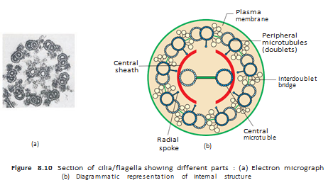
Centrosome and Centrioles
Centrosome — An organelle usually containing two centrioles surrounded by amorphous pericentriolar material.
- Orientation: The two centrioles lie perpendicular to each other; each has a cartwheel-like organisation.
- Centriole structure: Nine evenly spaced peripheral tubulin fibrils, each a triplet, form the centriole; adjacent triplets are linked.
- Hub and spokes: The proteinaceous hub in the proximal region connects to peripheral triplets by protein radial spokes.
- Functions: Centrioles form ciliary and flagellar basal bodies and help form spindle fibres in animal-cell division.
Nucleus
Nucleus — A membrane-enclosed organelle containing chromatin that helps control cellular activities and heredity.
- Early descriptions: Robert Brown described the nucleus in 1831; Flemming named the basic-dye-stained nuclear material chromatin.
- Interphase nucleus: When the cell is not dividing, it contains chromatin, nucleoplasm (nuclear matrix) and one or more nucleoli.
- Nuclear envelope: Two parallel membranes separated by a – perinuclear space; it separates nuclear contents from the cytoplasm.
- Outer membrane: Usually continuous with the endoplasmic reticulum and often bears ribosomes.
- Nuclear pores: Form where the two envelope membranes fuse; allow RNA and protein molecules to move in both directions between nucleus and cytoplasm.
- Nucleolus: A non-membranous, spherical region where ribosomal RNA is actively synthesised; it is often larger and more numerous in cells with high protein synthesis.
- Nuclei per cell: Usually one, though the number varies. Mature mammalian erythrocytes and plant sieve-tube cells lack a nucleus.
- Chromatin: Contains DNA, histone proteins, non-histone proteins and RNA; it becomes organised into visible chromosomes during cell division.
- DNA quantity: A human cell contains about of DNA distributed among chromosomes ( pairs).
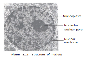
Chromosomes and Centromere Types
Chromosome — A structured form of chromatin visible during cell division, with a primary constriction called the centromere.
- Centromere: Holds the two chromatids of a chromosome; disc-shaped kinetochores occur on its sides.
- Classification: Chromosomes are classified by centromere position and the relative lengths of their arms.
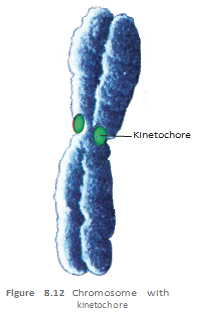
| Type | Centromere position | Arms |
|---|---|---|
| Metacentric | Middle | Two equal arms |
| Submetacentric | Slightly away from the middle | One shorter and one longer arm |
| Acrocentric | Close to one end | One extremely short and one very long arm |
| Telocentric | At the terminal end | Centromere is terminal |
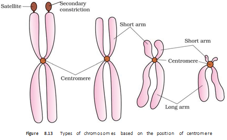
Secondary constriction
- Satellite: A chromosome may have a non-staining secondary constriction at a constant position, making a small fragment-like region appear.
Microbodies
Microbodies — Minute, membrane-bound vesicles containing various enzymes.
- Occurrence: Present in both plant and animal cells.
Why are mitochondria called the powerhouses of the cell?
State the axonemal microtubule arrangement in a typical eukaryotic cilium or flagellum.
Name the chromosome types based on centromere position.
Quick recap
- Mitochondria are double-membrane organelles; their inner cristae increase surface area for aerobic respiration and ATP production.
- Plastids include chloroplasts, chromoplasts and leucoplasts; chloroplast thylakoids contain chlorophyll.
- Light reactions occur in the grana, while dark reactions occur in the stroma.
- Ribosomes are non-membranous RNA–protein structures; eukaryotic ribosomes are and prokaryotic ribosomes are .
- The cytoskeleton supports cell shape and movement; cilia and flagella typically have a axoneme.
- Centrioles help form basal bodies and, in animal cells, spindle fibres during cell division.
- The nucleus contains chromatin and nucleoli; chromosomes are classified by centromere position, and microbodies are enzyme-containing vesicles found in plant and animal cells.
Biology — ALL CHAPTERS
1
The Living World
2
Biological Classification
3
Plant Kingdom
4
Animal Kingdom
5
Morphology of Flowering Plants
6
Anatomy Of Flowering Plants
7
Structural Organization In Animals
8
Cell The Unit Of Life
9
Biomolecules
10
Cell Cycle And Cell Division
11
Photosynthesis in Higher Plants
12
Respiration In Plants
13
Plant Growth And Development
14
Breathing And Exchange Of Gases
15
Body Fluids And Circulation
16
Excretory Products And Their Elimination
17
Locomotion And Movement
18
Neural Control And Coordination
19
Chemical Coordination and integration