Class 11 PCB . Biology . Biology . Locomotion and Movement
Chapter 17: Locomotion And Movement
Ch 17
Biology
Class 11 PCB
Introduction
Movement and Locomotion
Movement — A change in position of an organism or a body part.
Locomotion — A voluntary movement that changes an organism’s place or location.
How movement and locomotion are related
- Examples of movement: Protoplasm streams in Amoeba; cilia, flagella and tentacles move; humans move limbs, jaws, eyelids and tongue.
- Examples of locomotion: Walking, running, climbing, flying and swimming change an organism’s location.
- Relationship: All locomotion is movement, but not all movement is locomotion.
- Shared structures: Paramoecium cilia move food and aid locomotion; Hydra tentacles capture prey and assist locomotion; human limbs support posture changes and locomotion.
- Why animals move: To find food, shelter, mates, breeding grounds or favourable conditions, and to escape predators. Locomotion varies with habitat and situation.
Types of Movement
| Type | How it occurs | Examples / function |
|---|---|---|
| Amoeboid | Pseudopodia form through streaming protoplasm; microfilaments also contribute. | Macrophages and blood leucocytes move this way; Amoeba is another example. |
| Ciliary | Coordinated beating of cilia on ciliated epithelium. | Moves dust and foreign particles from the trachea; helps ova pass through the female reproductive tract. |
| Muscular | Contraction of muscle tissue. | Moves limbs, jaws and tongue; supports locomotion and other body movements. |
- Cilia and flagella: These are outgrowths of the cell membrane.
- Flagellar movement: Helps spermatozoa swim, maintains water currents in sponge canal systems, and aids locomotion in protozoans such as Euglena.
- Coordination: Locomotion depends on coordinated activity of the muscular, skeletal and nervous systems.
Muscle Tissue and Its Types
Muscle — A specialised tissue of mesodermal origin with properties that enable movement.
- Proportion: Muscles contribute about 40–50% of an adult human’s body weight.
- Properties: Muscles show excitability, contractility, extensibility and elasticity.
- Classification: Muscles are classified by location, appearance and regulation of activity; by location, they are skeletal, visceral or cardiac.
| Type | Location / appearance | Control and main role |
|---|---|---|
| Skeletal (striated, voluntary) | Associated with skeletal components; striped appearance under a microscope. | Voluntary control; mainly responsible for locomotion and changes in posture. |
| Visceral (smooth, nonstriated, involuntary) | Walls of hollow organs, such as the alimentary canal and reproductive tract; no striations. | Involuntary; transports food through the digestive tract and gametes through the genital tract. |
| Cardiac (striated, involuntary) | Heart; cells form a branching arrangement. | Involuntary; nervous system does not directly control its activity. |
Skeletal Muscle Structure
Organisation of skeletal muscle
- Whole muscle, held together by a common collagenous connective-tissue layer called fascia
- Muscle bundles (fascicles)
- Muscle fibres
- Parallel myofibrils (myofilaments) within each fibre
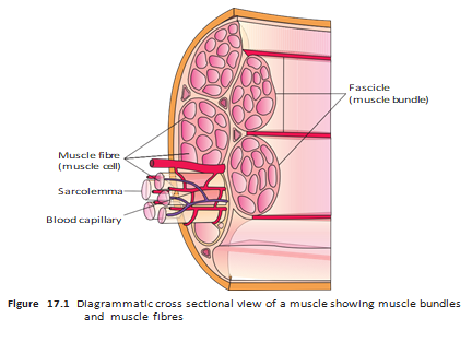
Muscle fibre
- Sarcolemma: The plasma membrane surrounding a muscle fibre; it encloses the sarcoplasm.
- Syncytium: A muscle fibre has many nuclei within its sarcoplasm.
- Sarcoplasmic reticulum: Specialised endoplasmic reticulum that stores calcium ions.
- Myofibrils: Numerous parallel contractile structures in the sarcoplasm, made of repeating thin and thick filaments.
| Structure | Composition / position |
|---|---|
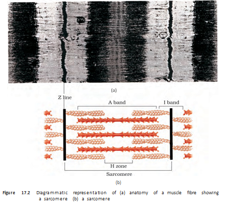
| I-band (isotropic band) | Light band containing thin actin filaments; a Z-line bisects each I-band. |
| A-band (anisotropic band) | Dark band containing thick myosin filaments. |
| Z-line | Elastic line at the centre of an I-band; thin filaments attach firmly to it. |
| M-line | Thin fibrous membrane at the middle of the A-band; holds thick filaments together. |
| H-zone | Central part of the thick filaments in a resting sarcomere that is not overlapped by thin filaments. |
Sarcomere and filament arrangement
- Striations: Alternating dark A-bands and light I-bands create the striped appearance of skeletal muscle.
- Filaments: Actin forms the thin filaments; myosin forms the thick filaments. Both run parallel to one another and to the myofibril’s length.
- Sarcomere: The portion of a myofibril between two successive Z-lines; it is the functional unit of muscle contraction.
- Resting arrangement: Thin filaments partially overlap the ends of thick filaments, leaving the central H-zone uncovered.
How are movement and locomotion related?
Name the three types of movement shown by human body cells and give one example of each.
What is a sarcomere, and how is it bounded?
Quick recap
- Locomotion changes location; all locomotion is movement, but many movements do not cause locomotion.
- Human cells show amoeboid, ciliary and muscular movements; cilia and flagella are cell-membrane outgrowths.
- Locomotion requires coordinated muscular, skeletal and nervous system activity.
- Muscles are mesodermal tissues with excitability, contractility, extensibility and elasticity.
- Skeletal, visceral and cardiac muscles differ in location, appearance and control.
- Myofibrils contain thin actin and thick myosin filaments arranged in alternating bands.
- The sarcomere, from Z-line to Z-line, is the functional unit of contraction.
Introduction Structure of Contractile Proteins
Structure of Contractile Proteins
Myosin filament — The thick filament is a polymer made of many myosin monomers called meromyosins.
Sliding-Filament Mechanism
Cross-Bridge Cycle and Contraction
- Calcium binds troponin → tropomyosin shifts, exposing myosin-binding sites on actin.
- Myosin binds actin → ATP hydrolysis provides energy for cross-bridge formation.
- Myosin head pivots → attached actin is pulled towards the centre of the band.
- A new ATP binds myosin → the cross-bridge breaks; ATP hydrolysis resets the myosin head for another cycle.
- Cycle repeats → thin filaments slide further until calcium is pumped back into the sarcoplasmic cisternae.
Sarcomere Changes and Relaxation
- Contraction: Actin pulls its attached lines inward, shortening the sarcomere.
- Band changes: The bands become shorter, while the band retains its length.
- Relaxation: Calcium returns to the sarcoplasmic cisternae; troponin-tropomyosin again masks actin's binding sites and the lines return to their original positions.
- Repeat activation: The cross-bridge cycle continues while calcium remains available and ATP is supplied.
Muscle Fibre Types and Fatigue
| Feature | Red fibres | White fibres |
|---|---|---|
| Myoglobin | Abundant; gives a reddish appearance and stores oxygen | Low amount; fibres appear pale or whitish |
| Mitochondria | Numerous; use oxygen for ATP production | Few |
| Main energy process | Aerobic; suited to using stored oxygen | Anaerobic |
| Sarcoplasmic reticulum | Not specified as abundant | Present in greater amount |
Do not say that the thick and thin filaments shorten: they slide past each other, shortening the sarcomere; the band stays the same length while the band narrows.
What changes occur in the sarcomere during contraction?
How does calcium enable actin and myosin to interact?
Quick recap
- Thin filament: Two F-actin strands coil together; tropomyosin and troponin regulate myosin-binding sites.
- Thick filament: Meromyosins form the filament; projecting HMM heads bind actin and hydrolyse ATP.
- Contraction signal: Acetylcholine triggers a sarcolemma action potential and calcium release into the sarcoplasm.
- Cross-bridge cycle: Calcium exposes actin sites; myosin binds, pulls actin, detaches when ATP binds, and resets.
- Sarcomere: The lines move inward, the bands narrow and the band stays the same length.
- Relaxation and fibres: Calcium reuptake masks actin sites; red fibres are myoglobin- and mitochondria-rich, while white fibres rely mainly on anaerobic energy.
Skeletal System
Skeletal System: Composition and Divisions
| Division | Bones | Main parts |
|---|---|---|
| Axial skeleton | 80 | Skull, vertebral column, sternum and ribs |
| Appendicular skeleton | 126 | Limb bones and their girdles |
Axial Skeleton
Skull
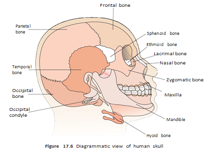
- Cranium: 8 bones form a hard protective covering around the brain.
- Facial bones: 14 bones form the front part of the skull; cranial and facial bones total 22.
- Hyoid: A single U-shaped bone at the base of the buccal cavity is also included in the skull.
- Ear ossicles: Each middle ear has three tiny bones—malleus, incus and stapes.
- Occipital condyles: Two condyles connect the skull to the upper vertebral column; this is a dicondylic skull.
Vertebral Column
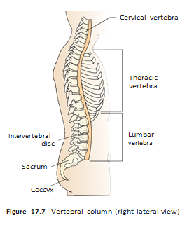
- Structure: A dorsal column of 26 serially arranged vertebral units, extending from the skull and forming the trunk’s main framework.
- Neural canal: The hollow central passage in each vertebra carries the spinal cord.
- Atlas: The first vertebra; it articulates with the skull’s occipital condyles.
- Functions: Protects the spinal cord, supports the head, and provides attachment for ribs and back muscles.
| Region (from skull downward) | Number of units |
|---|---|
| Cervical | 7 |
| Thoracic | 12 |
| Lumbar | 5 |
| Sacral | 1 fused unit |
| Coccygeal | 1 fused unit |
Sternum and Ribs
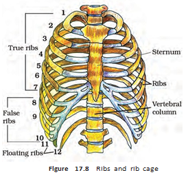
- Sternum: A flat bone along the ventral midline of the thorax.
- Ribs: There are 12 pairs; each is a thin, flat bone connected dorsally to the vertebral column.
- Bicephalic feature: Each rib has two articulation surfaces at its dorsal end.
- Rib cage: Thoracic vertebrae, ribs and sternum together form the rib cage.
| Type | Pairs | Connection to sternum |
|---|---|---|
| True ribs | 1st–7th | Directly connected by hyaline cartilage |
| Vertebrochondral (false) ribs | 8th–10th | Join the 7th rib through hyaline cartilage; no direct connection |
| Floating ribs | 11th–12th | Not connected ventrally |
Appendicular Skeleton
Appendicular skeleton — The bones of the limbs and their girdles, which connect the limbs to the axial skeleton.
| Limb | Bones |
|---|---|
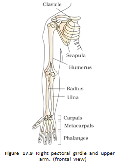
| Forelimb | Humerus; radius and ulna; 8 carpals; 5 metacarpals; 14 phalanges |
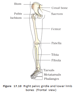
| Hindlimb | Femur; tibia and fibula; 7 tarsals; 5 metatarsals; 14 phalanges; patella at the knee |
Limb Bone Details
- Each limb: Contains 30 bones.
- Hand: Carpals form the wrist, metacarpals the palm, and phalanges the digits.
- Foot: Tarsals form the ankle, metatarsals the foot, and phalanges the digits.
- Femur: The thigh bone and the longest bone in the body.
- Patella: A cup-shaped kneecap that covers the knee on its ventral side.
Limb Girdles
Pectoral Girdle
- Structure: Two halves; each half contains a clavicle and a scapula.
- Scapula: A large, flat, triangular bone on the dorsal thorax, between the 2nd and 7th ribs.
- Spine and acromion: The scapular spine projects into the expanded acromion; the clavicle articulates with the acromion.
- Glenoid cavity: A depression below the acromion that receives the head of the humerus to form the shoulder joint.
- Clavicle: A long, slender, double-curved bone commonly called the collar bone.
Pelvic Girdle
- Structure: Two coxal bones form the pelvic girdle.
- Coxal bone: Each is formed by fusion of the ilium, ischium and pubis.
- Acetabulum: The cavity where these three bones meet; it receives the thigh bone.
- Pubic symphysis: The two halves meet ventrally at this joint, which contains fibrous cartilage.
Joints
Joint — A point of contact between bones, or between a bone and cartilage, through which muscle-generated force produces movement; the joint acts as a fulcrum.
| Type | Structure | Movement | Example |
|---|---|---|---|
| Fibrous | Bones joined by dense fibrous connective tissue, often as sutures | No movement | Flat bones of the skull |
| Cartilaginous | Bones joined by cartilage | Limited movement | Adjacent vertebrae |
| Synovial | A fluid-filled synovial cavity lies between the articulating bone surfaces | Considerable movement | Shoulder, knee, atlas–axis, carpals, thumb joint |
| Joint type | Example |
|---|---|
| Ball and socket | Humerus and pectoral girdle |
| Hinge | Knee |
| Pivot | Atlas and axis |
| Gliding | Between carpals |
| Saddle | Between a carpal and the thumb metacarpal |
Disorders of the Muscular and Skeletal System
| Disorder | Main feature or cause |
|---|---|
| Myasthenia gravis | Autoimmune disorder of the neuromuscular junction; causes skeletal-muscle fatigue, weakness and possible paralysis |
| Muscular dystrophy | Progressive degeneration of skeletal muscles, mostly due to genetic disorders |
| Tetany | Rapid muscle spasms caused by low in body fluids |
| Arthritis | Inflammation of joints |
| Osteoporosis | Age-related decrease in bone mass that raises fracture risk; decreased estrogen is a common cause |
| Gout | Joint inflammation due to uric acid crystal accumulation |
Name the two divisions of the human skeleton and their main components.
Distinguish true, vertebrochondral and floating ribs.
How do fibrous, cartilaginous and synovial joints differ in movement?
Quick recap
- The human skeleton has 206 bones and is divided into axial and appendicular parts.
- The axial skeleton includes the skull, vertebral column, sternum and 12 pairs of ribs.
- Each limb has 30 bones; girdles connect the limbs to the axial skeleton.
- Joints may be fibrous, cartilaginous or synovial, with increasing movement between these types.
- Myasthenia gravis, muscular dystrophy, tetany, arthritis, osteoporosis and gout affect muscles, bones or joints.
Biology — ALL CHAPTERS
1
The Living World
2
Biological Classification
3
Plant Kingdom
4
Animal Kingdom
5
Morphology of Flowering Plants
6
Anatomy Of Flowering Plants
7
Structural Organization In Animals
8
Cell The Unit Of Life
9
Biomolecules
10
Cell Cycle And Cell Division
11
Photosynthesis in Higher Plants
12
Respiration In Plants
13
Plant Growth And Development
14
Breathing And Exchange Of Gases
15
Body Fluids And Circulation
16
Excretory Products And Their Elimination
17
Locomotion And Movement
18
Neural Control And Coordination
19
Chemical Coordination and integration