Class 11 PCB . Biology . Biology . Excretory Products and Their Elimination
Chapter 16: Excretory Products And Their Elimination
Ch 16
Biology
Class 11 PCB
Introduction
Excretory Products and Nitrogenous Wastes
| Waste and process | Animals | Elimination and water balance |
|---|---|---|
| Ammonia — ammonotelism | Many bony fishes, aquatic amphibians and aquatic insects | Readily soluble; usually diffuses across body surfaces or fish gills as ammonium ions. Kidneys have little role. Highly toxic and requires much water. |
| Urea — ureotelism | Mammals, many terrestrial amphibians and marine fishes | Ammonia is converted to urea in the liver; blood carries it to the kidneys for filtration and excretion. Some urea may be retained in the kidney matrix to help maintain osmolarity. |
| Uric acid — uricotelism | Reptiles, birds, land snails and insects | Excreted as a pellet or paste, with minimal water loss; it is the least toxic of the three wastes. |
Terrestrial Adaptation and Water Conservation
- Terrestrial life increases the need to conserve water.
- Animals produce less-toxic nitrogenous wastes such as urea or uric acid instead of relying on highly toxic ammonia.
- Uric acid can be eliminated with especially little water loss.
Excretory Structures in Animals
| Structure | Animals | Main role |
|---|---|---|
| Protonephridia (flame cells) | Platyhelminthes, including Planaria; rotifers; some annelids; Amphioxus | Mainly regulate ions and fluid volume (osmoregulation). |
| Nephridia | Earthworms and other annelids | Remove nitrogenous wastes and maintain fluid and ionic balance. |
| Malpighian tubules | Most insects, including cockroaches | Remove nitrogenous wastes and support osmoregulation. |
| Antennal glands (green glands) | Crustaceans, such as prawns | Perform excretory functions. |
Human Excretory System
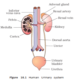
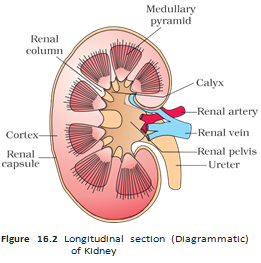
Internal Structure of the Kidney
Nephron: The Functional Unit
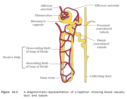
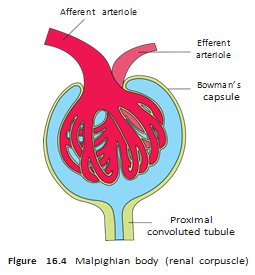
Nephron Types and Blood Supply
| Feature | Cortical nephron | Juxtamedullary nephron |
|---|---|---|
| Loop of Henle | Short; extends only slightly into the medulla. | Long; runs deep into the medulla. |
| Vasa recta | Absent or highly reduced. | Present as vessels running parallel to the long loop. |
Compare ammonotelic, ureotelic and uricotelic animals by their nitrogenous waste and water loss.
Name the two main parts of a nephron and the structure formed by the glomerulus enclosed by Bowman’s capsule.
What structures make up the human excretory system?
Quick recap
- Animals eliminate nitrogenous wastes, carbon dioxide, water and excess ions.
- Ammonia is most toxic and needs much water; uric acid is least toxic and conserves water.
- Terrestrial adaptation favored less-toxic urea and uric acid to reduce water loss.
- Animal excretory structures include protonephridia, nephridia, Malpighian tubules and antennal glands.
- The human excretory system consists of kidneys, ureters, urinary bladder and urethra.
- Each kidney has nearly one million nephrons; the nephron includes a glomerulus and renal tubule.
- Cortical nephrons have short loops of Henle; juxtamedullary nephrons have long loops extending deep into the medulla.
Urine Formation
The Three Processes of Urine Formation
Overview
- Urine formation occurs in the nephron through glomerular filtration, tubular reabsorption and tubular secretion.
- Filtration moves substances from blood into Bowman’s capsule; reabsorption returns useful substances to blood; secretion adds selected substances to the filtrate.
- Overall balance: about litres of filtrate are formed daily, but only about litres of urine are released.
Glomerular Filtration and GFR
Glomerular filtration — Pressure-driven filtration of blood through the glomerulus into the lumen of Bowman’s capsule.
Filtration Barrier
- Driving force: glomerular capillary blood pressure pushes blood plasma through three layers.
- Layers: endothelium of glomerular blood vessels → basement membrane → epithelium of Bowman’s capsule.
- Podocytes: epithelial cells of Bowman’s capsule have filtration slits (slit pores) between their extensions.
- Ultrafiltration: nearly all plasma constituents pass into Bowman’s capsule, while plasma proteins are mostly retained in the blood.
- Renal blood flow: kidneys filter about –, roughly one-fifth of the blood pumped by each ventricle per minute.
Glomerular filtration rate (GFR) — The volume of filtrate formed by both kidneys per unit time.
GFR Regulation
- Normal GFR: about , or approximately , in a healthy person.
- Juxtaglomerular apparatus (JGA): a sensitive region formed where the distal convoluted tubule (DCT) contacts the afferent arteriole.
- Response to falling GFR: JG cells release renin, which stimulates glomerular blood flow and helps restore GFR towards normal.
Reabsorption and Secretion
How Substances Move Across Tubules
- Reabsorption: nearly of the filtrate is reclaimed by renal tubules, reducing about of filtrate to roughly of urine.
- Active reabsorption: glucose, amino acids and are taken back from the filtrate using energy.
- Passive reabsorption: nitrogenous wastes are reabsorbed by passive transport; water also moves passively in the nephron’s initial segments.
- Tubular secretion: tubular cells transfer , and ammonia into the filtrate.
- Why secretion matters: it helps maintain ionic and acid–base balance in body fluids.
Functions of the Nephron Tubules
| Segment | Main features and functions |
|---|---|
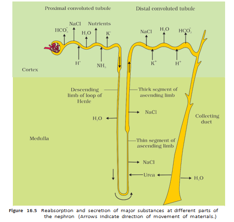
| Proximal convoluted tubule (PCT) | Simple cuboidal brush-border epithelium increases surface area. Reabsorbs nearly all essential nutrients and about – of electrolytes and water. Secretes and ammonia; reabsorbs to help maintain pH and ionic balance. |
| Descending limb of Henle’s loop | Permeable to water but almost impermeable to electrolytes; water leaves the filtrate, concentrating it as it descends. |
| Ascending limb of Henle’s loop | Impermeable to water; electrolytes move out actively or passively. The filtrate becomes more dilute as it ascends; reabsorption here is minimal. |
| Distal convoluted tubule (DCT) | Conditionally reabsorbs and water; can reabsorb . Secretes selected , and to regulate pH and sodium–potassium balance. |
| Collecting duct | Allows substantial water reabsorption, concentrating urine. Small amounts of urea pass into the medullary interstitium to support its osmolarity; secretion of and helps maintain pH and ionic balance. |
Concentration of the Filtrate
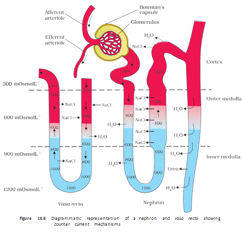
Counter-current mechanism — Opposite-direction flows in the limbs of Henle’s loop and vasa recta, together with their close arrangement, establish and maintain the medullary osmotic gradient.
Medullary Osmotic Gradient
- Opposite flows: filtrate moves in opposite directions in the two limbs of Henle’s loop; blood also flows in opposite directions through the two limbs of the vasa recta.
- Gradient: osmolarity rises from about in the cortex to about in the inner medulla.
- Main contributors: and urea build and maintain the medullary interstitial gradient.
- recycling: salt leaves the ascending limb of Henle’s loop, exchanges with the descending vasa recta, and is returned to the interstitium from the ascending vasa recta.
- Urea recycling: small amounts of urea enter the thin ascending limb of Henle’s loop and are transported back into the interstitium from the collecting tubule.
- Result: the interstitial gradient draws water from the collecting tubule, concentrating the urine; human kidneys can produce urine nearly four times as concentrated as the initial filtrate.
How the Counter-current System Concentrates Urine
- Opposite flow in Henle’s loop and vasa recta supports exchange of and urea.
- The exchange maintains high osmolarity in the inner medullary interstitium.
- Water moves out of the collecting tubule into the concentrated interstitium.
- The filtrate becomes concentrated urine.
What is GFR, and how is a fall in GFR corrected?
How does the counter-current mechanism help produce concentrated urine?
Quick recap
- Urine formation consists of glomerular filtration, tubular reabsorption and tubular secretion.
- The filtration barrier passes most plasma constituents but retains most proteins; healthy GFR is about .
- The JGA helps regulate GFR: falling GFR triggers renin release from JG cells.
- Tubule segments selectively reabsorb water and useful substances and secrete ions to regulate body-fluid balance.
- Counter-current exchange maintains a medullary osmotic gradient from about to .
- The medullary gradient promotes water reabsorption and allows urine to become nearly four times as concentrated as the initial filtrate.
Regulation Of Kidney Function
Hormonal Regulation of Kidney Function
Osmoreceptors and ADH
- Osmoreceptors detect changes in blood volume, body-fluid volume and ionic concentration.
- Low fluid volume: Activates osmoreceptors, prompting the hypothalamus to release antidiuretic hormone (ADH, or vasopressin) from the neurohypophysis.
- ADH: Increases water reabsorption in later parts of the nephron, reducing urine output and conserving water.
- Feedback: Restored fluid volume switches off osmoreceptor stimulation and suppresses ADH release.
- Blood vessels: ADH also constricts blood vessels, raising blood pressure; this can increase glomerular blood flow and GFR.
Renin–Angiotensin Mechanism
- A fall in glomerular blood flow, glomerular blood pressure or GFR activates juxtaglomerular (JG) cells.
- JG cells release renin; it converts blood angiotensinogen to angiotensin I, which is further converted to angiotensin II.
- Angiotensin II constricts blood vessels, raising glomerular blood pressure and GFR.
- Angiotensin II stimulates the adrenal cortex to release aldosterone.
- Aldosterone increases sodium and water reabsorption in the distal tubule, raising blood pressure and GFR.
Renin–angiotensin mechanism — A hormonal feedback mechanism that helps restore glomerular blood pressure and GFR while increasing sodium and water reabsorption.
Atrial Natriuretic Factor (ANF)
- Trigger: Increased blood flow to the heart’s atria causes release of ANF.
- Effect: ANF dilates blood vessels and lowers blood pressure.
- Role: ANF acts as a check on the renin–angiotensin mechanism.
Micturition
Micturition — The process of releasing urine from the urinary bladder through the urethra.
Micturition Reflex
- Urine is stored in the urinary bladder until a voluntary signal is given by the central nervous system (CNS); filling stretches the bladder.
- Stretch receptors in the bladder wall send signals to the CNS.
- The CNS sends motor messages that contract the bladder’s smooth muscles and relax the urethral sphincter.
- Urine is released; the neural mechanism coordinating this response is the micturition reflex.
Urine: Normal Features and Clinical Significance
- Average output: An adult excretes about – of urine and – of urea per day.
- Appearance: Urine is light-yellow and watery, has a characteristic odour and is slightly acidic, with .
- Clinical use: Urine analysis can help diagnose metabolic disorders and kidney malfunction.
- Glycosuria: Glucose in urine; ketonuria: ketone bodies in urine. Both can indicate diabetes mellitus.
Other Organs Involved in Excretion
| Organ | Substances eliminated | Key point |
|---|---|---|
| Lungs | Large amounts of carbon dioxide and some water | Remove about of carbon dioxide per minute. |
| Liver | Bilirubin, biliverdin, cholesterol, degraded steroid hormones, vitamins and drugs | These are secreted in bile; most ultimately leave with digestive wastes. |
| Sweat glands in skin | Water, , small amounts of urea and lactic acid | Sweat mainly cools the body surface but also removes some wastes. |
| Sebaceous glands in skin | Sterols, hydrocarbons and waxes | These leave in sebum, which forms a protective oily covering on the skin. |
| Salivary glands | Small amounts of nitrogenous wastes | Saliva provides a minor route of waste elimination. |
Disorders of the Excretory System
Uremia — A harmful condition in which kidney malfunction causes urea to accumulate in the blood; it can lead to kidney failure.
Hemodialysis
- Blood is drawn from an artery and mixed with an anticoagulant such as heparin.
- Blood passes through a coiled cellophane tube inside a dialysing unit.
- The surrounding dialysing fluid resembles plasma but lacks nitrogenous wastes; these wastes diffuse through the porous membrane down their concentration gradient.
- Cleared blood is given anti-heparin and returned to the body through a vein.
Kidney Transplantation
- Use: Kidney transplantation is the ultimate method for correcting acute renal failure.
- Donor: A functioning kidney is transplanted, preferably from a close relative, to reduce the chance of immune rejection.
- Outcome: Modern clinical procedures have improved the success rate of this complex treatment.
- Renal calculi
- Stones or insoluble masses of crystallised salts, such as oxalates, formed within the kidney.
- Glomerulonephritis
- Inflammation of the kidney glomeruli.
How do ADH and the renin–angiotensin mechanism help regulate kidney function?
What is the micturition reflex?
How does hemodialysis remove nitrogenous wastes from blood?
Quick recap
- Osmoreceptors and ADH help conserve water; ADH also constricts blood vessels and can raise GFR.
- The renin–angiotensin mechanism restores glomerular pressure and GFR; ANF lowers blood pressure and checks this mechanism.
- Urine is stored in the bladder until a voluntary CNS signal; bladder stretch initiates the micturition reflex.
- Urine analysis can reveal glycosuria or ketonuria, which may indicate diabetes mellitus.
- Lungs, liver, skin and salivary glands also eliminate some wastes.
- Uremia may be treated with hemodialysis; transplantation is the ultimate treatment for acute renal failure.
Biology — ALL CHAPTERS
1
The Living World
2
Biological Classification
3
Plant Kingdom
4
Animal Kingdom
5
Morphology of Flowering Plants
6
Anatomy Of Flowering Plants
7
Structural Organization In Animals
8
Cell The Unit Of Life
9
Biomolecules
10
Cell Cycle And Cell Division
11
Photosynthesis in Higher Plants
12
Respiration In Plants
13
Plant Growth And Development
14
Breathing And Exchange Of Gases
15
Body Fluids And Circulation
16
Excretory Products And Their Elimination
17
Locomotion And Movement
18
Neural Control And Coordination
19
Chemical Coordination and integration