Class 11 PCB . Biology . Biology . Body Fluids and Circulation
Chapter 15: Body Fluids And Circulation
Ch 15
Biology
Class 11 PCB
Introduction
Transport in Animals
- Transport: Animals move nutrients, oxygen and other essential substances to cells and carry wastes away.
- Mechanisms: Simple animals use water in body cavities, while complex animals use body fluids such as blood and lymph.
Blood
- Blood: A fluid connective tissue that transports substances throughout the body.
- Components: Blood consists of plasma and formed elements, including red blood cells, white blood cells and platelets.
Plasma
- Plasma: The straw-coloured fluid matrix of blood, making up about 55% of its volume.
- Composition: It contains mostly water, along with proteins, mineral ions and dissolved nutrients.
- Functions: Plasma proteins support clotting, defence and osmotic balance; plasma without clotting factors is called serum.
Formed Elements
- Formed elements: The cellular components of blood—erythrocytes, leucocytes and platelets—make up about 45% of blood.
- Roles: These components transport respiratory gases, defend the body and help blood clot.
Erythrocytes (Red Blood Cells)
- Erythrocytes: Red blood cells are the most abundant blood cells and contain haemoglobin, which transports respiratory gases.
- Structure: In mammals, mature RBCs are generally biconcave and lack a nucleus.
- Life span: RBCs live for about 120 days and are destroyed in the spleen.
Leucocytes (White Blood Cells)
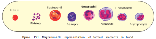
Platelets (Thrombocytes)
Differentiate plasma and serum.
Name the two groups of WBCs and their members.
Quick recap
- Blood is a connective tissue with plasma () and formed elements ().
- Plasma contains water, proteins, dissolved substances and inactive clotting factors; plasma without clotting factors is serum.
- RBCs contain haemoglobin, transport respiratory gases and are destroyed in the spleen after about days.
- WBCs are nucleated defence cells classified as granulocytes or agranulocytes.
- Platelets are megakaryocyte-derived cell fragments essential for clotting; low counts can cause excessive bleeding.
Blood Groups
ABO Blood Grouping
ABO donor patterns
- Group O has no A or B antigens on RBCs, so it can donate to all ABO groups; it is called the universal donor in the ABO system.
- Group AB has no anti-A or anti-B antibodies in its plasma, so it can receive from all ABO groups; it is called the universal recipient in the ABO system.
- Compatibility check: The donor’s RBC antigens must not react with the recipient’s plasma antibodies.
Rh Blood Grouping
Erythroblastosis foetalis — A potentially fatal condition in which maternal anti-Rh antibodies destroy the RBCs of an Rh+ foetus, possibly causing severe anaemia and jaundice.
Preventing Rh incompatibility
- Prevention: Anti-Rh antibodies are administered to an Rh− mother immediately after the delivery of her first child to prevent sensitisation.
Coagulation of Blood
Main steps in clot formation
- Injury activates platelets and clotting factors.
- Thrombokinase complex helps convert inactive prothrombin into thrombin.
- Thrombin converts soluble plasma fibrinogen into fibrin threads.
- Fibrin network traps blood cells and forms the clot.
Lymph (Tissue Fluid)
Functions of lymph
- Fluid return: The lymphatic system collects tissue fluid and drains it back into major veins.
- Immunity: Specialised lymphocytes in lymph help produce immune responses.
- Transport: Lymph carries nutrients and hormones.
- Fat absorption: Fats are absorbed through lymph in the lacteals of intestinal villi.
How can an Rh− mother carrying an Rh+ foetus develop erythroblastosis foetalis?
Describe the main sequence of blood clot formation.
State whether compatibility refers to ABO or Rh grouping: the universal donor/recipient labels here describe the ABO system, while Rh matching is an additional requirement.
Quick recap
- ABO groups depend on A and B antigens on RBCs and corresponding antibodies in plasma.
- Group O is the ABO universal donor; group AB is the ABO universal recipient.
- Rh+ blood has the Rh antigen; Rh− blood lacks it and can form anti-Rh antibodies after exposure.
- Maternal anti-Rh antibodies can destroy an Rh+ foetus’s RBCs in a later pregnancy; anti-Rh antibodies after first delivery help prevent this.
- Thrombin converts fibrinogen into fibrin, which forms the clotting network.
- Lymph returns tissue fluid to major veins, supports immunity, transports substances and absorbs fats through intestinal lacteals.
Circulatory Pathways
Types of Circulatory Pathways
- Circulatory patterns: Animals have either open or closed circulatory systems.
- Open system: Blood enters open spaces or body cavities; a closed system keeps blood within vessels.
- Regulation: A closed system allows blood flow to be controlled more precisely.
Vertebrate Hearts and Circulation
Blood flow in the main vertebrate patterns
- Fish: heart → gills (oxygenation) → body → heart.
- Amphibians/reptiles: oxygenated blood enters the left atrium and deoxygenated blood enters the right atrium → both enter one ventricle and mix.
- Birds/mammals/crocodiles: right heart → lungs; left heart → body, with no mixing between the streams.
Human Circulatory System
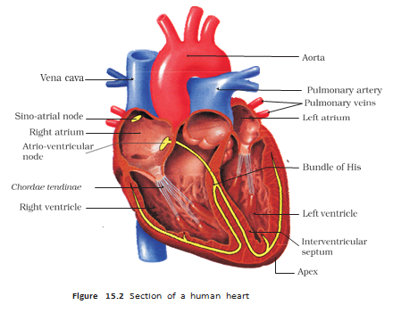
| Structure | Position or opening | Function |
|---|---|---|
| Interatrial septum | Between the right and left atria; thin and muscular | Separates the atria |
| Interventricular septum | Between the ventricles; thick-walled | Separates the ventricles |
| Atrioventricular septum | Between each atrium and ventricle; fibrous tissue with an opening | Connects chambers on the same side through the guarded opening |
| Tricuspid valve | Right atrium → right ventricle | Three cusps; prevents backward flow |
| Bicuspid (mitral) valve | Left atrium → left ventricle | Prevents backward flow |
| Semilunar valves | Right ventricle → pulmonary artery; left ventricle → aorta | Prevent blood from flowing back into the ventricles |
Heart's Nodal Tissue and Conduction Pathway
- Nodal tissue: Specialised cardiac muscle that generates action potentials without external stimulation; it is autoexcitable.
- SAN (sinoatrial node): Located in the upper-right corner of the right atrium; generates the highest rate, about – action potentials per minute.
- Pacemaker: The SAN initiates and maintains rhythmic heart contractions; the normal heart rate averages about beats per minute.
- AVN (atrioventricular node): Located in the lower-left region of the right atrium near the atrioventricular septum.
- Conduction route: SAN → AVN → AV bundle (bundle of His) → right and left branches → Purkinje fibres through the ventricles.
Cardiac Cycle
Cycle of Heart Contraction and Relaxation
- Joint diastole: chambers fill → atrial systole completes ventricular filling.
- Ventricular systole: atrioventricular valves close → semilunar valves open → blood is ejected.
- Ventricular diastole: semilunar valves close → atrioventricular valves open → chambers fill again.
At about beats per minute, one cardiac cycle lasts approximately seconds.
Stroke volume is the blood pumped by one ventricle in one cycle. Cardiac output is the volume pumped by each ventricle per minute; it averages about (or ) in a healthy person.
Heart Sounds
- First sound (lub): Closure of the tricuspid and bicuspid valves.
- Second sound (dub): Closure of the semilunar valves.
- Importance: These sounds can be heard with a stethoscope and have clinical diagnostic value.
Electrocardiogram (ECG)
Why is the SAN called the pacemaker?
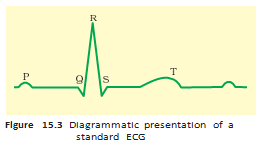
State the meaning of the P wave, QRS complex and T wave in an ECG.
What are stroke volume and cardiac output?
Quick recap
- Circulation may be open or closed; closed vessels allow more precise regulation of blood flow.
- Fish have single circulation, amphibians and most reptiles have incomplete double circulation, and birds, mammals and crocodiles have double circulation.
- The human heart has four chambers, valves for one-way flow, and a conduction system led by the SAN.
- A cardiac cycle alternates chamber systole and diastole; each ventricle ejects about per beat.
- Cardiac output equals stroke volume multiplied by heart rate and averages about per minute.
- ECG waves indicate electrical events: P for atria, QRS for ventricles, and T for ventricular recovery.
Double Circulation
Blood Vessels
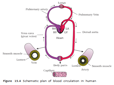
- Blood vessels: These form a branching network that carries blood between the heart and body tissues.
- Arteries and veins: Arteries carry blood away from the heart, while veins return it to the heart.
- Capillaries: Their thin walls allow exchange of substances between blood and tissues.
Pulmonary and Systemic Circulation
Special Circulatory Pathways
- Special pathways: Some blood vessels provide dedicated routes for blood flow to particular organs.
- Organ supply: These pathways support the circulation needs of organs such as the liver and heart.
Regulation of Cardiac Activity
Use this formula to calculate the volume pumped by one ventricle per minute.
Disorders of the Circulatory System
Hypertension — Hypertension is blood pressure persistently above normal; repeated readings of or higher indicate hypertension.
Coronary artery disease (CAD) — CAD, often referred to as atherosclerosis, affects the vessels supplying blood to the heart muscle.
Angina (angina pectoris) — Angina is acute chest pain that occurs when the heart muscle does not receive enough oxygen.
Heart failure — Heart failure is the inability of the heart to pump blood effectively enough to meet the body's needs.
Why is human circulation called double circulation?
How do autonomic signals affect cardiac activity and cardiac output?
Quick recap
- Double circulation has pulmonary and systemic pathways; blood passes through the heart twice during one complete circulation.
- Pulmonary circulation takes blood from the right ventricle to the lungs and returns it to the left atrium.
- Systemic circulation sends blood from the left ventricle to body tissues and returns it to the right atrium.
- The hepatic portal vein carries blood from the intestine to the liver; coronary vessels serve the heart muscle.
- The myogenic heart is moderated by ANS signals and adrenal medullary hormones; sympathetic activity increases and parasympathetic activity decreases cardiac output.
- Cardiac output equals stroke volume multiplied by heart rate and is approximately .
- Hypertension, CAD, angina and heart failure affect blood pressure, coronary blood flow or the heart's pumping ability.
Biology — ALL CHAPTERS
1
The Living World
2
Biological Classification
3
Plant Kingdom
4
Animal Kingdom
5
Morphology of Flowering Plants
6
Anatomy Of Flowering Plants
7
Structural Organization In Animals
8
Cell The Unit Of Life
9
Biomolecules
10
Cell Cycle And Cell Division
11
Photosynthesis in Higher Plants
12
Respiration In Plants
13
Plant Growth And Development
14
Breathing And Exchange Of Gases
15
Body Fluids And Circulation
16
Excretory Products And Their Elimination
17
Locomotion And Movement
18
Neural Control And Coordination
19
Chemical Coordination and integration