Class 11 PCB . Biology . Biology . Neural Control and Coordination
Chapter 18: Neural Control And Coordination
Ch 18
Biology
Class 11 PCB
Introduction
Coordination and Integration
Coordination — The process by which two or more organs interact and complement one another’s functions to help maintain homeostasis.
| System | How it coordinates |
|---|---|
| Neural system | Provides organised point-to-point connections for rapid coordination. |
| Endocrine system | Provides chemical integration through hormones. |
Coordination During Exercise
- Exercise begins → muscular activity and energy demand increase.
- Oxygen demand rises → respiration rate, heartbeat and blood flow increase.
- Exercise stops → nerves, lungs, heart, kidneys and other organs gradually return to normal.
Neural System in Animals
Neuron — A highly specialised cell that detects, receives and transmits different kinds of stimuli.
| Animal group / example | Neural organisation |
|---|---|
| Hydra | A simple network of neurons. |
| Insects | A brain with several ganglia and neural tissues. |
| Vertebrates | A more highly developed neural system. |
Human Neural System
| Division | Components / function |
|---|---|
| Central neural system (CNS) | Brain and spinal cord; processes information and controls responses. |
| Peripheral neural system (PNS) | All nerves of the body associated with the brain and spinal cord. |
| Point | Afferent fibres | Efferent fibres |
|---|---|---|
| Direction of impulses | From tissues and organs to the CNS. | From the CNS to the relevant peripheral tissues and organs. |
| Role | Carry incoming information to the CNS. | Carry regulatory impulses from the CNS. |
| Division | Main function |
|---|---|
| Somatic neural system | Relays impulses from the CNS to skeletal muscles. |
| Autonomic neural system | Carries impulses from the CNS to involuntary organs and smooth muscles. |
Autonomic and Visceral Neural Systems
- Autonomic divisions: The autonomic neural system is further divided into the sympathetic and parasympathetic neural systems.
- Visceral nervous system: The PNS complex of nerves, fibres, ganglia and plexuses that carries impulses between the CNS and the viscera.
Neuron: Structural and Functional Unit
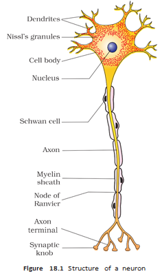
| Part | Structure and function |
|---|---|
| Cell body | Contains cytoplasm, typical cell organelles and Nissl’s granules. |
| Dendrites | Short, repeatedly branched fibres containing Nissl’s granules; carry impulses towards the cell body. |
| Axon | Long fibre that carries impulses away from the cell body towards a synapse or neuromuscular junction. |
| Synaptic knobs | Bulb-like endings of axon branches containing synaptic vesicles with neurotransmitters. |
| Type | Structure | Example / location |
|---|---|---|
| Multipolar | One axon and two or more dendrites. | Cerebral cortex. |
| Bipolar | One axon and one dendrite. | Retina of the eye. |
| Unipolar | Cell body with one axon only. | Usually found in the embryonic stage. |
| Point | Myelinated axon | Nonmyelinated axon |
|---|---|---|
| Schwann cells | Envelop the nerve fibre and form a myelin sheath around the axon. | Enclose the nerve fibre but do not form a myelin sheath around the axon. |
| Distinct feature | Gaps between adjacent myelin sheaths are called nodes of Ranvier. | Myelin sheath and nodes of Ranvier are absent. |
| Distribution | Found in spinal and cranial nerves. | Common in autonomic and somatic neural systems. |
Distinguish between afferent and efferent nerve fibres.
Name the three neuron types based on the number of axons and dendrites, and give one location for each.
Quick recap
- Coordination lets organs work together to maintain homeostasis; neural and endocrine systems provide rapid point-to-point and hormonal integration, respectively.
- The CNS consists of the brain and spinal cord and processes information; the PNS consists of associated body nerves.
- Afferent fibres carry impulses to the CNS, while efferent fibres carry impulses away from it.
- The PNS includes somatic and autonomic divisions; the autonomic division includes sympathetic and parasympathetic systems.
- A neuron has a cell body, dendrites and an axon; dendrites carry impulses towards the cell body and axons carry them away.
- Neurons are multipolar, bipolar or unipolar; axons may be myelinated or nonmyelinated.
Generation and Conduction of Nerve Impulse
Resting Polarisation of the Axon and Resting Potential
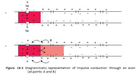
- Resting axon: The resting axonal membrane is more permeable to K⁺ than to Na⁺ and is impermeable to negatively charged proteins.
- Ion distribution: The axoplasm has high K⁺ and low Na⁺ concentrations, while the fluid outside has high Na⁺ and low K⁺ concentrations.
- Resting potential: The sodium-potassium pump moves 3 Na⁺ out for every 2 K⁺ moved in, maintaining a negatively charged inner surface and a positively charged outer surface.
Generation and Conduction of a Nerve Impulse
Transmission of Impulses Across Synapses
| Electrical synapse | Chemical synapse | |
|---|---|---|
| Connection | Pre- and post-synaptic membranes are in very close proximity. | The membranes are separated by a fluid-filled synaptic cleft. |
| Transmission | Electrical current passes directly from one neuron to the other. | Neurotransmitters carry the signal across the cleft. |
| Speed and occurrence | Faster than chemical transmission; rare in our nervous system. | Slower than electrical transmission. |
Chemical Synapse: Signal Transfer
Chemical Synapse Transmission
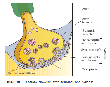
- Impulse arrives at the pre-synaptic axon terminal.
- Vesicles move and fuse with the pre-synaptic membrane, releasing neurotransmitters into the synaptic cleft.
- Neurotransmitters bind to specific receptors on the post-synaptic membrane.
- Ion channels open; ion entry may produce an excitatory or inhibitory potential in the post-synaptic neuron.
How is a nerve impulse generated and conducted along an axon?
How does a chemical synapse transmit an impulse?
Quick recap
- Resting axon: Neurons are excitable because their membranes are polarised; the inside is negative relative to the outside.
- Resting potential: The electrical potential difference across the resting axonal membrane; ion gradients are maintained in part by the sodium-potassium pump.
- Action potential: Sodium influx depolarises the membrane; local currents carry the impulse along the axon.
- Recovery: Increased potassium permeability and outward diffusion help restore resting potential.
- Electrical synapse: Direct current transfer between closely placed membranes; faster and rare.
- Chemical synapse: Neurotransmitters cross the cleft and can produce an excitatory or inhibitory post-synaptic potential.
Central Neural System
Brain: Control Centre and Protection
Neural System and Signalling
- Role: The neural system coordinates and integrates body functions, including the metabolic and homeostatic activities of organs.
- Divisions: The human neural system consists of the central neural system (CNS) and peripheral neural system.
- CNS: Consists of the brain and spinal cord.
- Neurons: These functional units are excitable cells because ions are present at different concentrations across their membranes.
- Resting potential: The electrical potential difference across a neural membrane when it is at rest.
- Impulse conduction: A nerve impulse travels along the axon membrane as a wave of depolarisation followed by repolarisation.
- Synapse: The junction formed by the membranes of a pre-synaptic and a post-synaptic neuron; the membranes may be separated by a synaptic cleft.
- Chemical synapse: Neurotransmitters are the chemicals that transmit impulses across chemical synapses.
Major Brain Divisions
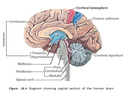
- Three regions: The brain is divided into the forebrain, midbrain and hindbrain.
Forebrain
Main Parts
- Components: The forebrain consists of the cerebrum, thalamus and hypothalamus.
- Cerebrum: The largest part of the human brain; a deep longitudinal cleft divides it into left and right cerebral hemispheres.
- Corpus callosum: A tract of nerve fibres connecting the two cerebral hemispheres.
- Thalamus: A major coordinating centre for sensory and motor signalling.
| Structure/area | Features and function |
|---|---|
| Cerebral cortex (grey matter) | Folded outer layer of the cerebral hemispheres; neuron cell bodies are concentrated here, giving it a grey appearance. |
| White matter | Inner part of the cerebral hemispheres; myelin-covered nerve fibres give it an opaque white appearance. |
| Motor areas | Control voluntary movement. |
| Sensory areas | Receive and process sensory information. |
| Association areas | Neither clearly sensory nor motor; support intersensory associations, memory and communication. |
Hypothalamus and Limbic System
- Hypothalamus: Lies at the base of the thalamus and contains centres regulating body temperature and the urge to eat and drink.
- Neurosecretory cells: Groups of these cells in the hypothalamus secrete hypothalamic hormones.
- Limbic system: Inner regions of the cerebral hemispheres and associated deep structures, including the amygdala and hippocampus, form this system.
- Functions: With the hypothalamus, it is involved in olfaction, autonomic responses, sexual behaviour, emotional reactions (excitement, pleasure, rage and fear) and motivation.
Midbrain
- Location: Between the thalamus/hypothalamus of the forebrain and the pons of the hindbrain.
- Cerebral aqueduct: A canal passing through the midbrain.
- Corpora quadrigemina: Four rounded swellings on the dorsal side of the midbrain.
- Sensory integration: The midbrain receives and integrates visual, tactile and auditory inputs.
Hindbrain and Brain Stem
| Structure | Key features and functions |
|---|---|
| Pons | Fibre tracts interconnect different regions of the brain. |
| Cerebellum | Its highly convoluted surface provides space for many neurons; it integrates information from the semicircular canals of the ear and the auditory system. |
| Medulla oblongata | Connected to the spinal cord; contains centres controlling respiration, cardiovascular reflexes and gastric secretions. |
Brain stem — The brain stem consists of the midbrain, pons and medulla oblongata, and forms the connections between the brain and spinal cord.
Quick recap
- The neural system coordinates and integrates organ functions; the CNS consists of the brain and spinal cord.
- Neurons are excitable due to ion concentration differences across their membranes; resting potential is the electrical difference across a resting membrane.
- Nerve impulses propagate as waves of depolarisation and repolarisation; chemical synapses use neurotransmitters.
- The brain is protected by the skull and meninges and controls body functions as well as higher mental activities.
- The forebrain includes the cerebrum, thalamus and hypothalamus; the hemispheres are joined by the corpus callosum.
- The midbrain integrates visual, tactile and auditory inputs; the hindbrain comprises the pons, cerebellum and medulla.
- The brain stem is made up of the midbrain, pons and medulla oblongata and connects the brain to the spinal cord.
Biology — ALL CHAPTERS
1
The Living World
2
Biological Classification
3
Plant Kingdom
4
Animal Kingdom
5
Morphology of Flowering Plants
6
Anatomy Of Flowering Plants
7
Structural Organization In Animals
8
Cell The Unit Of Life
9
Biomolecules
10
Cell Cycle And Cell Division
11
Photosynthesis in Higher Plants
12
Respiration In Plants
13
Plant Growth And Development
14
Breathing And Exchange Of Gases
15
Body Fluids And Circulation
16
Excretory Products And Their Elimination
17
Locomotion And Movement
18
Neural Control And Coordination
19
Chemical Coordination and integration