NEET UG . NEET Chemistry . Chemistry . Biomolecules
Chapter 19: Biomolecules
Ch 19
NEET Chemistry
NEET UG
Carbohydrates
Biomolecules and Carbohydrates
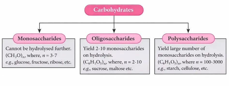
Reducing and Non-reducing Sugars
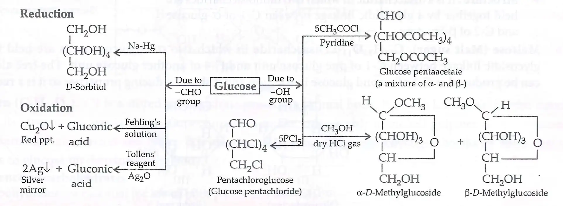
- Reducing sugars: Have a free aldehydic or ketonic group and reduce Fehling’s solution and Tollens’ reagent.
- Examples: Monosaccharides and disaccharides with a free reducing group, such as maltose and lactose, are reducing sugars.
- Non-reducing sugars: Lack a free aldehydic or ketonic group; sucrose is an example.
Monosaccharides and Glucose
Structure of Glucose
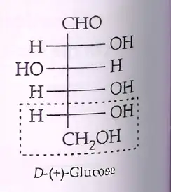
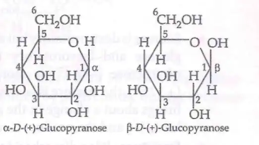
Glucose: Cyclic Forms and Anomers
- Ring formation: an group adds intramolecularly to the group, forming a cyclic hemiacetal.
- Pyranose: glucose forms a six-membered ring called the pyranose structure.
- Anomeric carbon: the former carbonyl carbon, -1, becomes a stereocentre; it is also called the glycosidic carbon.
- Anomers: α- and β-D-glucose differ only in configuration at the anomeric -1.
- Crystallisation: the α-form is obtained from a concentrated glucose solution at ; the β-form is obtained from a hot, saturated aqueous solution at .
Fructose
| Relationship | Meaning |
|---|---|
| Anomers | Differ in configuration at the anomeric carbon only. |
| Epimers | Differ in configuration at one chiral carbon only. |
| Enantiomers | Non-superimposable mirror-image stereoisomers; configuration differs at all stereocentres. |
| Diastereomers | Stereoisomers that are not mirror images. |
Disaccharides
Sucrose Hydrolysis and Invert Sugar
- Products: sucrose hydrolysis gives an equimolar mixture of D-(+)-glucose and D-(-)-fructose.
- Optical rotations: glucose rotates light by and fructose by .
- Invert sugar: the mixture is laevorotatory because fructose’s laevorotation is greater than glucose’s dextrorotation.
- Inversion: hydrolysis changes the sign of rotation from positive (dextro) to negative (laevo); the product mixture is called invert sugar.
Polysaccharides: Starch
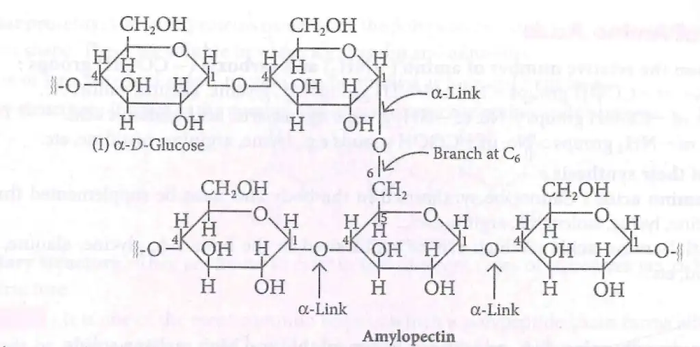
| Property | Amylose | Amylopectin |
|---|---|---|
| Structure | Long, unbranched chain | Branched chain |
| Linkages | α-1,4 | α-1,4 in chains; α-1,6 at branches |
| Solubility / starch proportion | Water-soluble; about 15–20% of starch | Branched component; proportion not specified |
Cellulose and Glycogen
- Cellulose: A linear condensation polymer of β-glucose units linked by β-glycosidic bonds between C-1 and C-4.
- Glycogen: The storage carbohydrate of animals, made of highly branched α-D-glucose units; it occurs in the liver, muscles and brain.
- Breakdown: Enzymes break glycogen down to glucose when the body needs it.
Importance of Carbohydrates
How are reducing and non-reducing sugars distinguished?
Why are α- and β-glucose called anomers?
What is invert sugar, and why is it laevorotatory?
Quick recap
- Carbohydrates are polyhydroxy aldehydes or ketones, or compounds that yield them on hydrolysis.
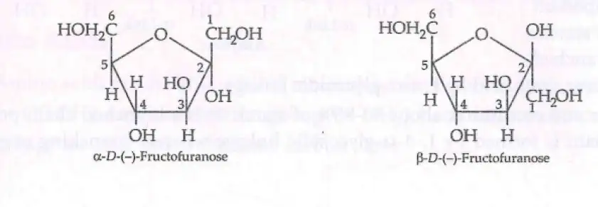
- Glucose is an aldohexose with α- and β-pyranose anomers at -1; fructose is a ketohexose with furanose anomers at -2.
- Sucrose is non-reducing; maltose and lactose are reducing disaccharides.
- Sucrose hydrolysis yields invert sugar, a laevorotatory mixture of glucose and fructose.
- Starch stores carbohydrate in plants, glycogen in animals, and cellulose has structural roles.
- Starch or cellulose hydrolysis gives glucose; cellulose also forms plant and bacterial cell walls, cotton and wood.
Proteins
Proteins and Amino Acids
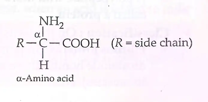
α-Amino acid — A bifunctional molecule with an amino group and a carboxyl group attached to the same α-carbon atom.
- General structure: The α-carbon is attached to , , hydrogen and a variable side chain .
- Side chain: The group differs among amino acids and gives each its distinct identity.
Classification of Amino Acids
| Type | Condition | Examples |
|---|---|---|
| Neutral | Number of groups = number of groups | Glycine, alanine, valine |
| Acidic | Number of groups > number of groups | Aspartic acid, glutamic acid |
| Basic | Number of groups > number of groups | Lysine, arginine, histidine |
| Type | Meaning | Examples |
|---|---|---|
| Essential | Cannot be synthesised by the body; must be obtained through the diet | Valine, leucine, lysine, isoleucine, arginine |
| Non-essential | Can be synthesised by the body | Glycine, alanine, glutamic acid, aspartic acid |
Properties of Amino Acids
- Physical properties: Usually colourless, crystalline, water-soluble solids with high melting points.
- Salt-like behaviour: Their acidic carboxyl group and basic amino group occur in the same molecule, so amino acids behave like salts.
- Reactions: They can form salts with both acids and bases; their reactions resemble those of primary amines and carboxylic acids.
Zwitterion — A dipolar ion formed in water when the carboxyl group loses a proton and the amino group accepts one; it has both positive and negative charges but is net neutral.
Isoelectric point — The pH at which an amino acid exists as a net-neutral zwitterion and does not migrate towards either electrode.
- At the isoelectric point: Positive and negative charges balance; amino acids have their least solubility in water, which helps in their separation.
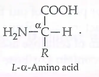
- Optical activity: All naturally occurring α-amino acids except glycine have a chiral asymmetric carbon and are optically active.
- L-configuration: In the conventional L-structure, the group is written on the left.
Structure of Proteins
Peptide bond — An amide linkage formed between the carboxyl group of one amino acid and the amino group of another, with loss of a water molecule.
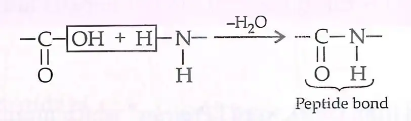
Peptides — Products formed by linking amino acids through peptide linkages.
| Name | Number of amino acid residues |
|---|---|
| Dipeptide | 2 |
| Tripeptide | 3 |
| Tetrapeptide | 4 |
| Oligopeptide | 2–10 |
| Polypeptide | More than 10 |
| Protein | A polypeptide with more than 100 residues and molecular mass above 10,000 u |
Classification by Molecular Structure
| Point | Fibrous proteins | Globular proteins |
|---|---|---|
| Shape and arrangement | Parallel polypeptide chains held together by hydrogen and disulphide bonds | Polypeptide chains coil into a compact, three-dimensional spherical shape |
| Solubility | Insoluble in water | Soluble in water |
| Examples | Keratin (hair, wool, silk); myosin (muscles) | Insulin; albumins |
Four Levels of Protein Structure
| Level | Meaning and key features |
|---|---|
| Primary | Number and linear sequence of amino acids joined by peptide bonds. |
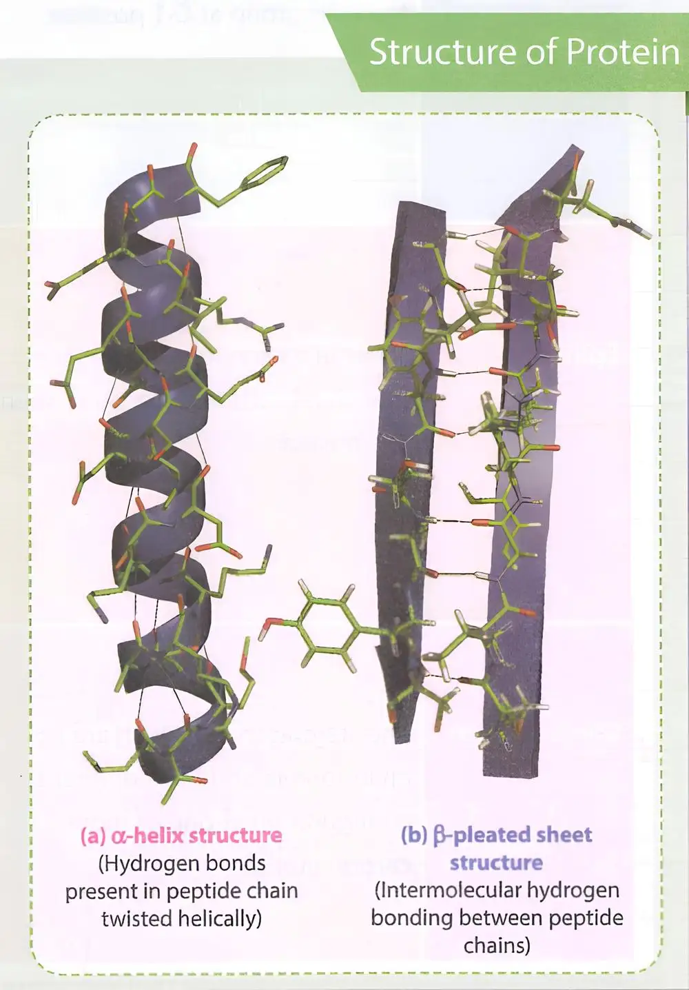
| Secondary | Local folding into α-helix or β-pleated sheet; stabilised by hydrogen bonds. |
| Tertiary | Overall folding of the polypeptide, including further folding of secondary structures; produces fibrous or globular forms. |
| Quaternary | Spatial arrangement of two or more polypeptide subunits in a protein. |
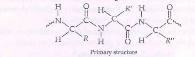
Secondary structures
- α-Helix: The chain twists into a right-handed screw; the group of each residue hydrogen-bonds to the group on an adjacent turn.
- β-Pleated sheet: Extended peptide chains lie side by side and are held together by intermolecular hydrogen bonds.
Tertiary structure: stabilising forces
- Forces: Hydrogen bonds, disulphide linkages, van der Waals forces and electrostatic attractions stabilise secondary and tertiary structures.
Denaturation of Proteins
Denaturation — Loss of a protein’s biological activity when physical or chemical changes disrupt its higher-level structure.
Denaturation pathway
- Change in temperature or pH → hydrogen bonds are disturbed
- Protein globules unfold and helices uncoil
- Biological activity is lost
- Secondary and tertiary structures change; the primary structure remains intact
- Examples: Boiling coagulates egg white; acid curdles milk, and cheese formation involves protein denaturation.
Biological Roles and Disease
- Defence: Some proteins act as antibodies and protect the body against foreign organisms.
- Sequence and disease: A change in the amino acid sequence of a protein can cause serious disease.
Sickle-cell anaemia
- In haemoglobin, glutamic acid is replaced by valine
- The amino acid sequence changes
- This altered haemoglobin causes sickle-cell anaemia
What is a peptide bond, and how is it formed?
What changes during protein denaturation, and what remains unchanged?
Quick recap
- Proteins are condensation polymers of α-amino acids linked by peptide bonds.
- Amino acids are classified by functional-group balance and whether the body can synthesise them.
- In water, amino acids can exist as zwitterions; least solubility occurs at the isoelectric point.
- Protein structure has four levels: primary, secondary, tertiary and quaternary.
- Fibrous proteins are generally water-insoluble; globular proteins are generally water-soluble.
- Denaturation disrupts secondary and tertiary structure but leaves the primary sequence intact.
- Replacing glutamic acid with valine in haemoglobin causes sickle-cell anaemia.
Enzymes
Enzymes: Biological Catalysts
Enzyme — A biological catalyst that speeds up biochemical reactions; almost all enzymes are globular proteins.
- Rate: Enzymes can greatly increase the rate of reactions.
- Specificity: They show high specificity for their reactants, called substrates, and for the products formed.
- Quantity: Enzymes are needed only in small quantities for a reaction to progress.
- Naming: Enzymes are generally named after the compound or class of compounds on which they act.
Mechanism of Enzyme Action
Activation energy — The energy barrier that must be overcome for a reaction to occur; enzymes lower this barrier, as chemical catalysts do.
- Effect: Lower activation energy allows a reaction to proceed more readily.
| Condition | Activation energy |
|---|---|
| Acid hydrolysis | |
| Enzyme-catalysed by sucrase |
Enzyme Deficiency and Disorders
Enzyme deficiency — A deficiency of a specific enzyme can cause a particular disorder or condition.
- Phenylalanine hydroxylase deficiency: Causes phenylketonuria.
- Tyrosinase deficiency: Causes albinism, a condition involving reduced melanin pigment in the skin.
Common Enzymes and Their Reactions
| Enzyme | Reaction catalysed |
|---|---|
| Amylase | Starch glucose |
| Maltase | Maltose glucose |
| Lactase | Lactose glucose + fructose |
| Pepsin | Proteins peptones |
| Trypsin | Proteins -amino acids |
| Nucleases | DNA, RNA nucleotides |
| Urease | Urea |
| Carbonic anhydrase |
How do enzymes increase the rate of biochemical reactions?
Name the enzyme deficiencies associated with phenylketonuria and albinism.
Quick recap
- Enzymes are biological catalysts; almost all are globular proteins.
- They act in small quantities, greatly increase reaction rates, and are highly specific for substrates and products.
- Enzymes generally take their names from the compounds or compound classes on which they act.
- Enzymes lower activation energy; sucrase lowers the activation energy of sucrose hydrolysis from to .
- Phenylalanine hydroxylase deficiency causes phenylketonuria; tyrosinase deficiency causes albinism.
- Know the enzyme–reaction pairs, including lactase: lactose to glucose and fructose.
Vitamins
Vitamins — Organic compounds needed in small amounts in the diet for specific functions and for normal growth and health.
Classification of Vitamins
| Point | Water-soluble | Fat-soluble |
|---|---|---|
| Solubility | Dissolve in water | Dissolve in fats and oils |
| Storage and excretion | Generally not stored; regularly excreted in urine. Vitamin B₁₂ is the exception to regular excretion. | Stored in the liver and adipose tissue |
| Vitamins | B₁, B₂, B₆, B₁₂ and C | A, D, E and K |
| Excess intake | Regular dietary supply is generally needed | Excess intake may cause hypervitaminosis |
Vitamin H
- Biotin: Vitamin H is listed as neither water-soluble nor fat-soluble in the source.
Vitamin Sources and Deficiency Diseases
| Vitamin | Chemical name | Solubility | Sources | Deficiency effects |
|---|---|---|---|---|
| A | Retinoids (retinol and carotenoids) | Fat | Milk, butter, eggs, fish, cod liver oil, green vegetables | Night-blindness; xerophthalmia |
| B₁ | Thiamine | Water | Pulses, nuts, cereals (rice, wheat), rice bran, yeast, egg yolk, fruits, green vegetables | Beri-beri; loss of appetite |
| B₂ | Riboflavin | Water | Milk, green vegetables, egg white, meat, liver, kidney | Anaemia; inflammation of tongue; dermatitis; cheilosis |
| B₆ | Pyridoxine | Water | Rice bran, whole cereals (wheat, gram), yeast, fish, meat, eggs | Central nervous system effects, weakness, convulsions, insomnia and anaemia |
| H | Biotin | Listed as neither water- nor fat-soluble | Milk, yeast, liver, kidney | Dermatitis |
| B₁₂ | Cyanocobalamin | Water | Eggs, milk, liver of ox or sheep, fish | Pernicious anaemia |
| C | Ascorbic acid | Water | Citrus fruits, chillies, sprouted pulses | Pyorrhea; scurvy (bleeding gums) |
| D | Ergocalciferol and cholecalciferol | Fat | Butter, milk, eggs, fish liver oil, liver, meat | Rickets (bending of bones); osteomalacia |
| E | Tocopherol | Fat | Wheat germ oil, milk, nuts, peanut oil, cottonseed oil, eggs, fish | Loss of fertility in males; muscle degeneration |
| K | Phylloquinone | Fat | Leafy vegetables such as cabbage and spinach | Haemorrhages; prolonged blood-clotting time |
Avitaminosis — A condition caused by vitamin deficiency.
Hypervitaminosis — A harmful condition associated with excess intake of fat-soluble vitamins.
Which vitamins are fat-soluble, and where are they stored?
What are avitaminosis and hypervitaminosis?
Quick recap
- Vitamins are dietary organic compounds needed in small amounts for normal growth and health.
- Water-soluble vitamins include B₁, B₂, B₆, B₁₂ and C; most are regularly excreted in urine.
- Fat-soluble vitamins A, D, E and K are stored in the liver and adipose tissue.
- Vitamin deficiency is called avitaminosis; excess fat-soluble vitamin intake may cause hypervitaminosis.
- Know the chemical names, dietary sources and deficiency effects of the listed vitamins.
Nucleic Acids
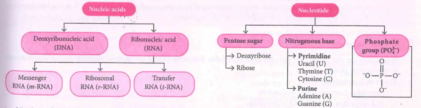
Nucleic acids — Polymers of nucleotides present in the nucleus of living cells; they transmit hereditary information and play an important role in protein synthesis.
Chemical Composition
Components of DNA and RNA
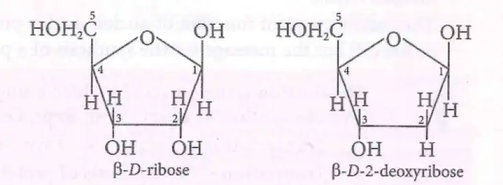
- Complete hydrolysis of DNA or RNA yields a pentose sugar, phosphoric acid and nitrogen-containing heterocyclic bases.
- DNA sugar: -D-2-deoxyribose; it lacks the -OH group present in ribose.
- RNA sugar: -D-ribose.
| Nucleic acid | Bases |
|---|---|
| DNA | Adenine (), guanine (), cytosine (), thymine () |
| RNA | Adenine (), guanine (), cytosine (), uracil () |
Nucleosides and Nucleotides
| Point | Nucleoside | Nucleotide |
|---|---|---|
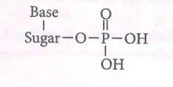
| Components | Pentose sugar + nitrogenous base | Pentose sugar + nitrogenous base + phosphate group |
| General structure | Sugar–base | Sugar–base–phosphate |
Structure of Nucleic Acids
| Level | Key features |
|---|---|
| Primary | Nucleotides are linked by phosphodiester linkages between the and carbon atoms of pentose sugars. |
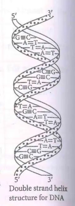
| Secondary: DNA | Two polynucleotide strands coil into a double helix. Sugar–phosphate backbones lie outside; bases face inward and pair through hydrogen bonds. |
| Secondary: RNA | A single strand folds back on itself to form a double helix. |
Complementary Base Pairing and Chargaff’s Rule
- DNA model: James Watson and Francis Crick gave the double-stranded helix structure for DNA.
- DNA base pairing: pairs with through 2 hydrogen bonds; pairs with through 3 hydrogen bonds.
- Chargaff’s rule: The amount of purines equals that of pyrimidines; in double-stranded DNA, and .
- RNA pairing: Adenine pairs with uracil, , through 2 hydrogen bonds.
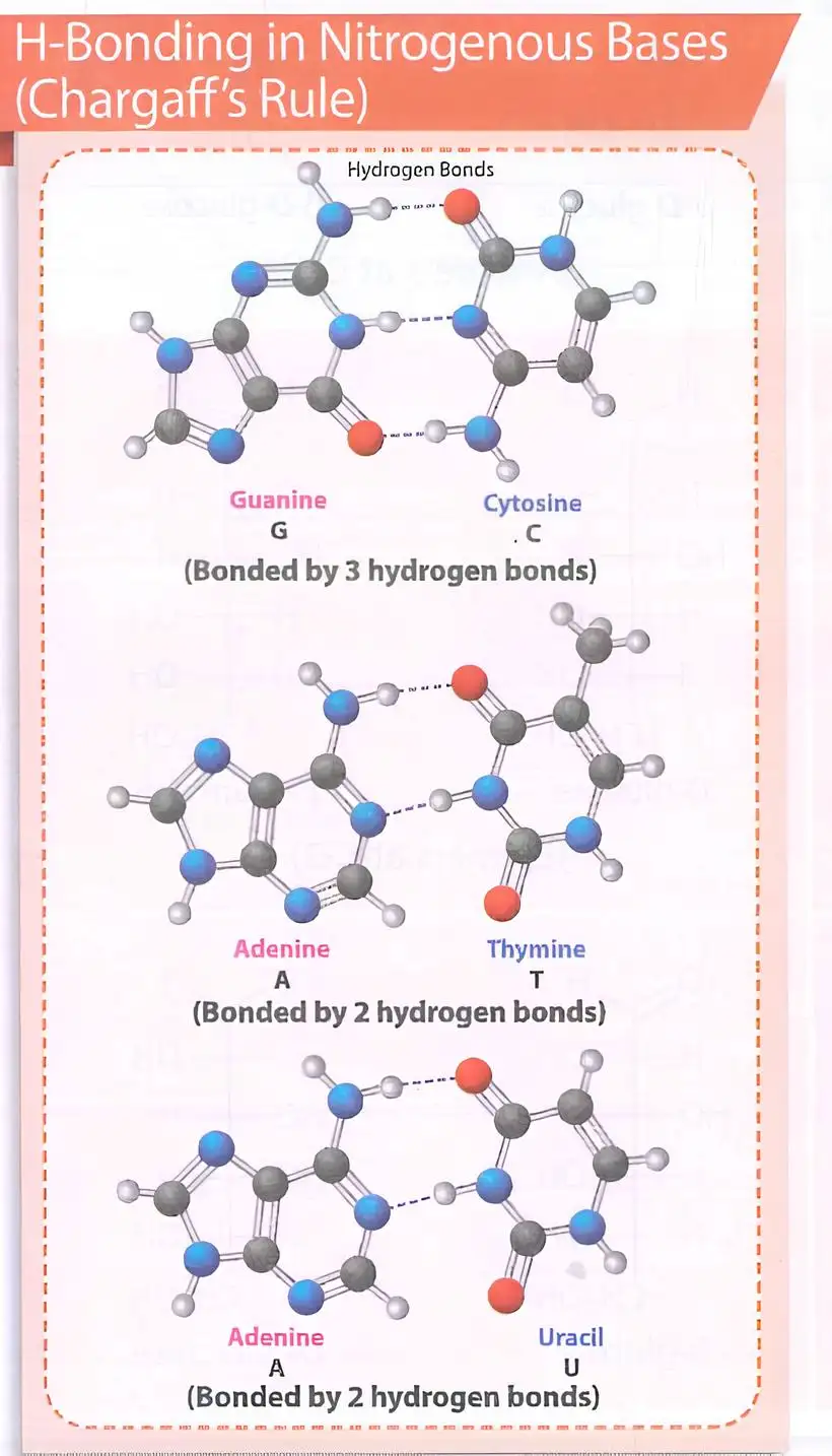
Do not confuse the stated pairs: DNA has pairing, while the text specifies pairing in RNA.
Biological Functions of Nucleic Acids
DNA and Heredity
- Genetic store: DNA stores genetic information and transmits hereditary characteristics.
- Species identity: DNA is exclusively responsible for maintaining the identity of different species.
- Replication: DNA can make two identical copies of itself; the copies are transferred to daughter cells during cell division.
- Protein instructions: DNA carries the message for synthesising specific proteins.
Replication — The process by which one DNA molecule produces two identical copies of itself.
Protein Synthesis
- Transcription: RNA is synthesised using the message in DNA.
- Translation: In the cytoplasm, mRNA directs protein synthesis with the help of rRNA and tRNA.
Genetic code — A linear sequence of nucleotide triplets in DNA or RNA that determines the specific amino-acid sequence during protein synthesis.
- Triplet code: Each code unit consists of 3 nucleotides.
- Significance: The genetic code is the biochemical basis of heredity and is nearly universal among organisms.
State Chargaff’s rule and the hydrogen-bonding pattern in DNA.
Differentiate a nucleoside from a nucleotide.
Quick recap
- Nucleic acids are nucleotide polymers involved in heredity and protein synthesis.
- DNA has deoxyribose and bases , , , and ; RNA has ribose and uses instead of .
- A nucleoside is sugar + base; a nucleotide is sugar + base + phosphate.
- DNA has a double helix; RNA has a single strand that can fold back to form a double helix.
- In DNA, forms 2 hydrogen bonds and forms 3; RNA pairing is also specified.
- DNA stores hereditary information, replicates, and supplies the message used in protein synthesis through transcription and translation.
- The genetic code consists of nucleotide triplets and is nearly universal.
Hormones
Hormones — Hormones are intercellular messenger molecules released directly into the bloodstream by endocrine glands.
| Type | Examples |
|---|---|
| Steroids | Estrogens and androgens |
| Polypeptides | Insulin and endorphins |
| Amino acid derivatives | Epinephrine and norepinephrine |
Roles of hormones
- Balance: Hormones help maintain the balance of biological activities in the body.
- Growth and development: Growth hormones and sex hormones contribute to these processes.
| Hormone | Effect on blood glucose |
|---|---|
| Insulin | Helps keep the blood glucose level within limits. |
| Glucagon | Tends to increase the blood glucose level. |
Blood glucose regulation
- Together: Insulin and glucagon regulate the blood glucose level.
Responses to external stimuli
- Epinephrine and norepinephrine: These hormones mediate responses to external stimuli.
Quick recap
- Hormones are intercellular messengers released into the bloodstream by endocrine glands.
- Hormone types include steroids, polypeptides, and amino acid derivatives.
- Hormones help maintain biological balance; growth and sex hormones support growth and development.
- Insulin helps keep blood glucose within limits, while glucagon tends to raise it; together they regulate blood glucose.
- Epinephrine and norepinephrine mediate responses to external stimuli.
NEET Chemistry — ALL CHAPTERS
1
Some Basic Concepts of Chemistry
2
Structure of Atom
3
Classification of Elements and Periodicity in Properties
4
Chemical Bonding and Molecular Structure
5
Thermodynamics
6
Equilibrium
7
Redox Reactions
8
Organic Chemistry - Some Basic Principles and Techniques
9
Hydrocarbons
10
Solutions
11
Electrochemistry
12
Chemical Kinetics
13
The d- and f- Block Elements
14
Coordination Compounds
15
Haloalkanes and Haloarenes
16
Alcohols, Phenols and Ethers
17
Aldehydes, Ketones and Carboxylic Acids
18
Amines
19
Biomolecules
20
The p-Block Elements (Group 13 and 14)
21
The p-Block Elements (Group 15 to 18)
22
Principles Related to Practical Chemistry